Infective Endocarditis
Infective endocarditis may be due to either pyogenic or opportunistic pathogens. In the latter case, they are often part of a systemic opportunistic infection with multiple organ localizations.
Fungal endocarditis has been reported with increasing frequency as the AIDS epidemic has gained momentum, helped by the compromise of cell-mediated immunity in patients with HIV infection [2, 3].Infective endocarditis occurs more frequently in intravenous drug users with AIDS, who comprise the second largest risk group for HIV infection after male homosexuals. These patients have frequent bacteremias, owing to the introduction of skin pathogens and talcum powder by unsterile intravenous injection, causing a higher risk of endocardial infection of right-sided cardiac valves. Infective endocarditis is higher in intravenous drug addicts who abuse multiple drugs (cocaine used intravenously in combination with heroin) in addition to alcohol.
The spectrum of pathogens responsible for endocardial infection in intravenous drug users with AIDS is not significantly different from that in HIV-uninfected drug users. However, owing to the deficit in cellular immunity, the pathogens are more virulent, leading to more significant cardiac structural damage and functional deterioration. Pyogenic bacteria more commonly causing infective endocarditis in AIDS are Staphylococcus aureus, Staphylococcus epi- dermidis, Streptococcus pneumoniae, and Haemophilus influenzae [2]. Infective endocarditis by Gram-negative bacteria, especially Pseudomonas species, has become more common in patients with AIDS, perhaps owing to the repeated hospitalizations that promote the acquisition of resistant organisms. Avirulent bacteria such as the HACEK group (Haemophilus species, Actinobacillus actinomycetemcomitans, Cardiobacterium hominis, Eikenella corrodens and Kingella kingae), which are often part of the endogenous flora of the mouth, can cause endocarditis in HIV-infected patients [2].
These bacteria are also difficult to culture from endocardial vegetations. Failure to obtain positive blood cultures in those patients with AIDS with strong clinical evidence for infective endocarditis should suggest prior antibiotic therapy or endocarditis by unusual bacteria (as well as HACEK organisms) or fungi.Fungal endocarditis, especially from Cryptococcus neoformans, Candida albicans, or Aspergillus fumigatus, is common in AIDS, particularly in intravenous drug abusers [2, 3]. It is generally related to systemic spread of fungal infection from extracardiac foci. Candidiasis of the oropharynx and esophagus is most often the primary focus, often progressing to systemic infection. Systemic cryptococcosis is one of the most common infections in AIDS patients. Although meningitis and encephalitis are the most frequent manifestations of cryptococcosis, cardiac involvement, particularly with pericardial effusion, is common [2]. Fungal myocarditis or myocardial abscesses may also occur in association with valve destruction [2].
Pathologic Features
Infective endocarditis is an ulcerative-polypous lesion due to a destructive valve process with thrombotic stratifications (Fig. 6). Thrombotic vegetations are usually gray, but their color is highly variable depending on the pathogen involved [2]. They are generally located on the endocardial surface of valve cusps but can be found also on mural endocardium. Their consistence is variable: they are friable at first and later become compact and adherent to the endocardium, owing to their organization. The friability is increased by lithic effects of bacteria and polymorphonuclear leukocytes [2, 3]. Valvular tissue destruction may involve the tensive apparatus with chordae tendinous rupture. Endocardial ulcerations at the cusp apposition lines are frequent, resulting in leaflets with a mouth-eaten look. On histology, thrombotic vegetations consist of fibrin and agglutinated platelets with inflammatory infiltration [3].
In the acute stage of endocardial infection, there is an infiltration with polymorphonuclear leukocytes with valve tissue necrosis; later there is a chronic inflammatory infiltration, made up of macrophages, lymphocytes and plasma cells, neoformed capillary vessels, and a fibroblastic proliferation that replaces the necrotic tissue and spreads at the base of thrombotic vegetation. In fungal endocarditis, however, the vegetations are made up essentially of fungal colonies without much fibrin and they may be so bulky that they obstruct the valve ostium [2, 3].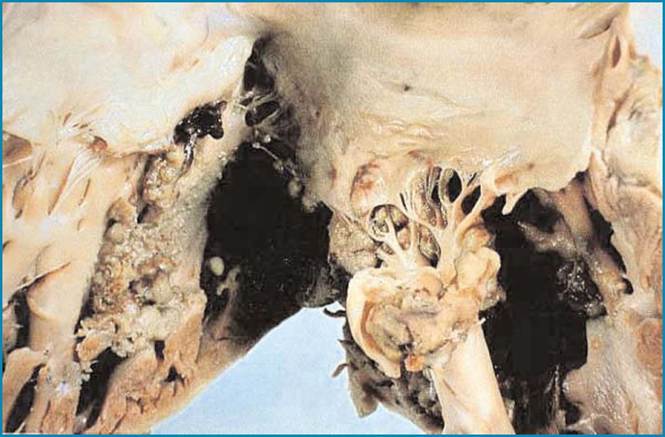
Fig. 6 Staphylococcus aureus endocarditis in an HIV-infected drug addicts who died of cardiogenic shock. The mitral valve shows numerous, large, grayish and friable vegetations. Involvement of the atrial endocardium is also shown. (Courtesy of Prof. D. Scevola, Department of Infectious and Parasitic Disease, University of Pavia, Italy)
When the left-side cardiac valves are involved, endocarditis can have a galloping course, with rapid onset of heart failure due to acute valvular insufficiency secondary to perforation of valve leaflets or a rupture of the tendinous chordae or papillary muscles [2]. Other complications are due to myocardial involvement with possible perforation of the ventricular septum or myocardial abscesses. The infection can extend to the pericardium with purulent pericarditis. The higher frequency of right-sided infectious endocarditis in HIV-infected intravenous drug users can explain the pulmonary embolic events with possible pulmonary cavitations and abscesses [2, 3]. The outcome is thrombus organization and fibrous repair. Residual bulky thrombotic polypi are often seen as calcific masses leaning out of both endocardial surfaces of the valvular leaflets [2].