Hyperthyroidism4,5
GENERAL PRINCIPLES
• Graves disease causes most cases of hyperthyroidism, especially in young patients. This autoimmune disorder may also cause proptosis (Graves orbitopathy) and pretibial myxedema, neither of which is found in other causes of hyperthyroidism.
• Toxic multinodular goiter (MNG) may cause hyperthyroidism, more commonly in older patients.
• Unusual causes include iodine-induced hyperthyroidism (precipitated by drugs such as amiodarone or radiographic contrast media), thyroid adenomas, subacute thyroiditis (painful tender goiter with transient hyperthyroidism), painless thyroiditis (nontender goiter with transient hyperthyroidism, most often seen in the postpartum period), and surreptitious ingestion of thyroid hormone. TSH-induced hyperthyroidism is extremely rare.
DIAGNOSIS
Clinical Presentation
HISTORY
• Symptoms include heat intolerance, weight loss, weakness, palpitations, oligomenorrhea, and anxiety.
• In the elderly, hyperthyroidism may present with only atrial fibrillation, heart failure, weakness, or weight loss, and a high index of suspicion is needed to make the diagnosis.
PHYSICAL EXAMINATION
• Signs include brisk tendon reflexes, fine tremor, proximal weakness, stare (related to eyelid retraction), and eyelid lag. Cardiac abnormalities may be prominent, including sinus tachycardia, atrial fibrillation, and exacerbation of coronary artery disease or heart failure.
• Key differentiating physical examination findings (Table 24-2) include the following:
î The presence of proptosis or pretibial myxedema, seen only in Graves disease (although many patients with Graves disease lack these signs)
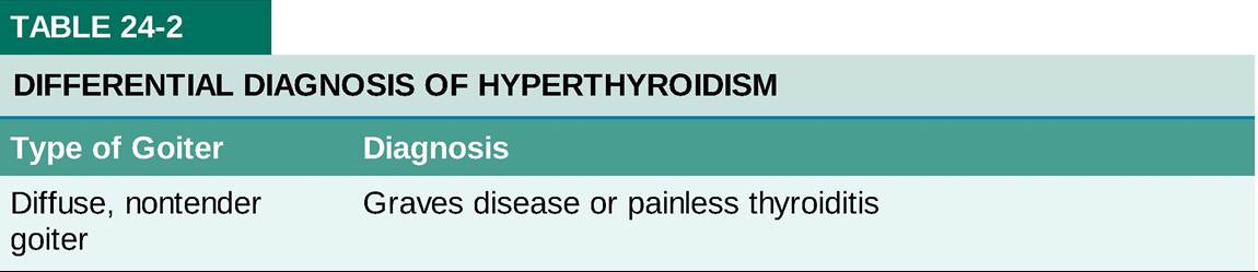
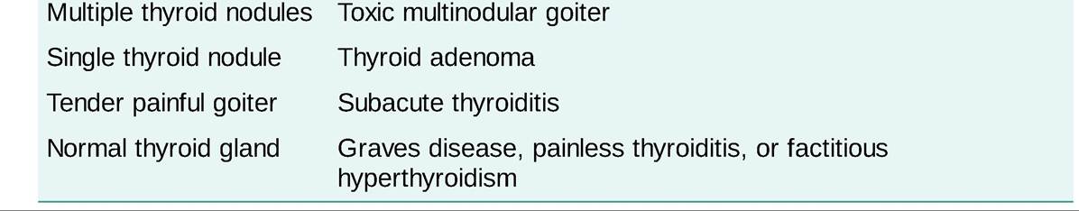
î A diffuse nontender goiter, consistent with Graves disease or painless thyroiditis
î Recent pregnancy, neck pain, or recent iodine administration, suggesting causes other than Graves
disease.
Diagnostic Testing
• In suspected hyperthyroidism, plasma TSH is the best initial diagnostic test.
î A normal TSH level virtually excludes clinical hyperthyroidism. If plasma TSH is low, plasma-free T4 should be measured to determine the severity of hyperthyroidism and as a baseline for therapy. If plasma-free T4 is elevated, the diagnosis of clinical hyperthyroidism is established.
î If plasma TSH is lt;0.1 #956;U#8725;mL but free T4 is normal, the patient may have clinical hyperthyroidism because of elevation of plasma T3 alone, and plasma-free T3 should be measured in this case.
î Mild (or subclinical) hyperthyroidism may suppress TSH to lt;0.1 #956;U#8725;mL, and thus suppression of TSH alone does not confirm that symptoms are due to hyperthyroidism.
î TSH may also be suppressed by severe nonthyroidal illness (see “Evaluation of Thyroid Function” section).
î In rare cases, 24-hour radioactive iodine uptake (RAIU) is needed to distinguish Graves disease or toxic nodules (in which RAIU is elevated) from postpartum thyroiditis, iodine-induced hyperthyroidism, or factitious hyperthyroidism (in which RAIU is very low).
TREATMENT
• Some forms of hyperthyroidism (subacute or postpartum thyroiditis) are transient and require only symptomatic therapy. A #946;-adrenergic antagonist (such as atenolol 25-100 mg daily) relieves symptoms of hyperthyroidism, such as palpitations, tremor, and anxiety, until hyperthyroidism is controlled by definitive therapy or until transient forms of hyperthyroidism subside. The dose is adjusted to alleviate symptoms and tachycardia and then reduced gradually as hyperthyroidism is controlled.
• Three methods are available for definitive therapy (none of which control hyperthyroidism rapidly): RAI, thionamides, and thyroidectomy.
î During treatment, patients are followed by clinical evaluation and measurement of plasma-free T4. Plasma TSH is not helpful in assessing the initial response to therapy because it remains suppressed until after the patient becomes euthyroid.
î Regardless of the therapy used, all patients with Graves disease require lifelong follow-up for recurrent hyperthyroidism or development of hypothyroidism.
• Choice of definitive therapy
î In Graves disease, RAI therapy is our preferred treatment for almost all patients. It is simple and highly effective but cannot be used in pregnancy (see “Hyperthyroidism” section). Thionamides achieve long-term control in fewer than half of patients with Graves disease, and they carry a small risk of life-threatening side effects (agranulocytosis, liver dysfunction). Thyroidectomy may be used in patients who refuse RAI therapy, have significant Graves ophthalmopathy, or who relapse or develop side effects with thionamide therapy.
î Other causes of hyperthyroidism: We prefer to treat toxic nodules with RAI (except in pregnancy or during breastfeeding), though thionamides or parti al thyroidectomy can also be used. Transient forms of hyperthyroidism because of thyroiditis should be treated symptomatically with atenolol. Iodine-induced hyperthyroidism is treated with thionamides and atenolol until the patient is euthyroid. Although treatment of some patients with amiodarone-induced hyperthyroidism with glucocorticoids has been advocated, nearly all patients with amiodarone-induced hyperthyroidism respond well to thionamide therapy.
• RAI therapy
î A single dose permanently controls hyperthyroidism in 90% of patients, and further doses can be given if necessary.
î A pregnancy test is done immediately before therapy in potentially fertile women.
î A 24-hour RAIU is usually measured and used to calculate the dose.
î Thionamides interfere with RAI therapy and should be stopped at least 3 days before treatment. If iodine treatment has been given, it should be stopped at least 2 weeks before RAI therapy.
î Follow-up: Usually, several months are needed to restore euthyroidism. Patients are evaluated at 4- to 6-week intervals, with assessment of clinical findings and plasma-free T4.
#9632; If thyroid function stabilizes within the normal range, the interval between follow-up visits is gradually increased to annual intervals.
#9632; If symptomatic hypothyroidism develops, thyroxine therapy is started (see “Hypothyroidism” section).
#9632; If symptomatic hyperthyroidism persists after 6 months, RAI treatment can be repeated.
î Side effects
#9632; Hypothyroidism occurs in most patients within the first year and thereafter continues to develop at a rate of approximately 3% per year.
#9632; Because of the release of stored hormone, a slight rise in plasma T4 may occur in the first 2 weeks after therapy. This development is important only in patients with severe cardiac disease, which may worsen as a result. Such patients should be treated with thionamides to restore euthyroidism and to deplete stored hormone before treatment with RAI.
#9632; There have been mixed results as to whether RAI has a clinically important effect on the course of Graves eye disease, but early treatment to prevent hypothyroidism after RAI seems to be beneficial. Patients should also be counseled to stop smoking, as this is a known contributor to worsening of the eye disease. Patients with clinical evidence of Graves orbitopathy should be evaluated by an ophthalmologist before tr eatment with RAI. Sometimes glucocorticoids or teprotumumab, an insulin-like growth factor 1 receptor antibody, is utilized in the treatment of Graves orbitopathy.
#9632; This treatment does not increase the risk of malignancy or cause congenital abnormalities in the offspring of women who conceive after RAI therapy.
• Thionamides: Methimazole and propylthiouracil (PTU) inhibit thyroid hormone synthesis. PTU also inhibits extrathyroidal deiodination of T4 to T3. Once thyroid hormone stores are depleted (after several weeks to months), T4 levels decrease. These drugs have no permanent effect on thyroid function. In the majority of patients with Graves disease, hyperthyroidism recurs within 6 months after therapy is stopped.
Spontaneous remission of Graves disease occurs in approximately one-third of patients during thionamide therapy, and in this minority, no other treatment may be needed. Remission is more likely in mild, recent onset hyperthyroidism and if the goiter is small. Because of a better safety profile, methimazole should be used instead of PTU except in specific situations (see the following text). î Initiation of therapy: Before starting therapy, patients must be warned of side effects and precautions. Usual starting doses are methimazole, 10-40 mg PO daily, or PTU, 100-200 mg PO tid; higher initial doses can be used in severe hyperthyroidism.î Follow-up: Restoration of euthyroidism takes up to several months.
#9632; Patients are evaluated at 4-week intervals with assessment of clinical findings and plasma-free T4. If plasma-free T4 levels do not fall after 4-8 weeks, the dose should be increased. Doses as high as methimazole, 60 mg PO daily, or PTU, 300 mg PO qid, may be required.
#9632; Once the plasma-free T4 level falls to normal, the dose is adjusted to maintain plasma-free T4 within the normal range.
#9632; No consensus exists on the optimal duration of therapy, but periods of 6 months to 2 years are usually used. Patients must be monitored carefully for recurrence of hyperthyroidism after the drug is stopped.
î Side effects are most likely to occur within the first few months of therapy.
#9632; Minor side effects include rash, urticaria, fever, arthralgias, and transient leukopenia.
#9632; Agranulocytosis occurs in 0.3% of patients treated with thionamides. Other life-threatening side effects include hepatitis, vasculitis, and drug-induced lupus erythematosus. These complications usually resolve if the drug is stopped promptly.
#9632; Patients must be warned to stop the drug immediately if jaundice or symptoms suggestive of agranulocytosis develop (e.g., fever, chills, sore throat) and to contact their physician promptly for evaluation.
Routine monitoring of the white blood cell count is not useful for detecting agranulocytosis, which develops suddenly.• Thyroidectomy: This procedure provides long-term control of hyperthyroidism in most patients.
î Surgery may trigger a perioperative exacerbation of hyperthyroidism, and patients should be prepared for surgery by one of two methods.
° A thionamide is given until the patient is nearly euthyroid. Supersaturated potassium iodide (SSKI), 40-80 mg (one to two drops) PO bid, is then added 1-2 weeks before surgery. Both drugs are stopped postoperatively.
î Atenolol (50-100 mg daily) is started 1-2 weeks before surgery. The dose of atenolol is increased, if necessary, to reduce the resting heart rate below 90 bpm and is continued for 5-7 days postoperatively. SSKI is given as mentioned earlier.
î Patients should be started on thyroxine replacement after surgery, and plasma TSH and free T4 should be measured 6 weeks later.
î Complications of thyroidectomy include hypothyroidism and hypoparathyroidism. Rare complications include permanent vocal cord paralysis, due to recurrent laryngeal nerve injury, and perioperative death.
SPECIAL CONSIDERATIONS
• Subclinical hyperthyroidism is present when the plasma TSH is below normal but the patient has no symptoms that are definitely caused by hyperthyroidism, and plasma levels of free T4 and T3 are normal.
î Subclinical hyperthyroidism increases the risk of atrial fibrillation in patients older than 65 years and those with heart disease and predisposes to osteoporosis in postmenopausal women; it should be treated in these patients. Treatment should also be considered in asymptomatic individuals without risk factors but with TSH persistently lt;0.1 #956;U#8725;mL.
î Asymptomatic young patients with mild Graves disease can be observed for spontaneous resolution of hyperthyroidism or the development of symptoms or increasing free T4 levels that warrant treatment.
• Urgent therapy is warranted when hyperthyroidism exacerbates heart failure or acute coronary syndromes and in rare patients with severe hyperthyroidism complicated by fever and delirium (thyroid storm). Concomitant diseases should be treated intensively, and confirmatory tests (serum TSH and free T4) should be obtained before therapy is started.
î PTU 300 mg PO q6h or methimazole 60 mg/d PO should be started immediately.
î Iodide (SSKI, two drops PO q12h) should be started to inhibit thyroid hormone secretion rapidly.
î Propranolol, 40 mg PO q6h (or an equivalent dose IV), should be given to patients with angina or myocardial infarction, and the dose should be adjusted to prevent tachycardia. #946;-Adrenergic antagonists may benefit some patients with heart failure and marked tachycardia but can further impair left ventricular systolic function. In patients with clinical heart failure, it should be given only with careful monitoring of left ventricular function.
î Plasma-free T4 is measured every 4-6 days. When free T4 approaches the normal range, the doses of methimazole and iodine are gradually decreased. RAI therapy can be scheduled 2-4 weeks after iodine is stopped.
• Hyperthyroidism in pregnancy: If hyperthyroidism is suspected, plasma TSH should be measured. Plasma TSH declines in early pregnancy but rarely to lt;0.1 #956;U#8725;mL due to the stimulatory effect of human chorionic gonadotropin on TSH receptors.
î If TSH is lt;0.1 #956;U#8725;mL, the diagnosis should be confirmed by measurement of plasma-free T4.
î RAI is contraindicated in pregnancy, and therefore, patients should be treated with PTU in the first trimester because of its lower risk of severe congenital defects, whereas methimazole can be used in later pregnancy. The dose should be adjusted at 4-week intervals to maintain the plasma-free T4 near the upper limit of the normal range to avoid fetal hypothyroidism. The dose required often decreases in the later stages of pregnancy.
î Atenolol, 25-50 mg PO daily, can be used to relieve symptoms while awaiting the effects of PTU.
î The fetus and neonate should be monitored for hyperthyroidism. The maternal plasma level of thyroid receptor antibodies should be assessed in early pregnancy, and if elevated or if the patient requires thionamide treatment during pregnancy, it should be repeated at weeks 18-22 and again at weeks 30-34 to assess this risk.