ACUTE LYMPHOBLASTIC LEUKEMIA (ALL)
Childhood ALL is considered as the prototype for cancer research, being the first disseminated malignancy cured by chemotherapy and irradiation. Currently, ~70-80% of these cases are potentially curable.
Epidemiology: Incidence of ALL is estimated to be ~3-10#8725;lac population; being 3-4 times more common in males. Peak age incidence is between 3-7 years.
Etiology: While exact etiology is not known, important hypotheses suggest:
• Genetic basis, due to higher risk in twins or siblings of index case (3-4 times) or in many genetic syndromes (10-20 times), e.g. Down syndrome, Bloom syndrome, Fanconi's anemia, neurofibromatosis and congenital immunodeficiency disorders, e.g. agammaglobulinemia or ataxia telangiectasia. Chromosomal abnormalities, e.g. aneuploidy or translocations are present in gt;80% of leukemic cells. Many germline single nucleotide polymorphisms have been identified with higher risk of ALL, involving genes like ARID5B, IKZF1, etc.
• Environmental basis, due to occurrence of many cases in geographical clusters or as second malignancy after radiotherapy or chemotherapy. Exposure to pesticides, pains and other chemicals as well as heavy paternal smoking in periconceptional period has also been implicated in higher risk of ALL.
Pathology: ALL is characterized by increased number of lymphoblasts in bone marrow with or without their presence in peripheral blood or infiltration to other organs.
A lymphoblast is characterized by:
• Typical morphology, i.e. high nucleus-cytoplasm ratio, dense nuclear chromatin and basophilic nongranular cytoplasm (Fig. 20.1),
• Cytochemical characteristics, i.e. presence of coarse granules/clumps on PAS staining, absence of peroxidase or Sudan-black positive granules and nonspecific esterase reaction,
• Biological markers, e.g. presence of terminal deoxynucleotidyl transferase (TdT) activity in B-progenitor and T-cell lymphoblasts, which is absent in normal lymphocytes.
Classification: ALL is classified according to lineage of blast cells and presence of immunological markers, as per WHO classification (Table 20.3). A small percentage of cases have markers for both lymphoid and myeloid derivation. In addition, chromosomal abnormalities are also used to classify the disease, for the purpose of treatment and outcome.
Clinical presentation: Initial symptoms are generally nonspecific and mimic a viral infection, e.g., fever,
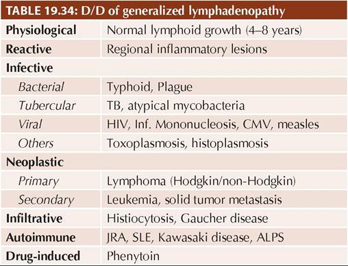
Fig. 20.1: (A) Normal lymphocyte; (B) Lymphoblasts.
TABLE 20.3: WHO classification of acute lymphoblastic leukemia
• B-Lymphoblastic leukemia (85%)
(earlier termed PreB or early PreB ALL)
• T-Lymphoblastic leukemia (15%)
(earlier termed T-cell ALL)
• Burkitt leukemia (lt;1%)
(arising from mature B cells)
anorexia and lethargy. Nearly 2/3rd of cases present within 4 weeks of onset of symptoms with:
• Signs of primary bone marrow failure—pallor, fever or persisting/recurrent infections (leukopenia) or mucocutaneous bleeding (thrombocytopenia)
• Signs of secondary organ infiltration—mild lymphadenopathy, splenohepatomegaly, bone/joint pains (due to perichondrial bone infiltration and subperiosteal bleeds) and headache/vomiting (due to meningeal involvement).
• Signs of mediastinal mass, e.g. SVC compression, dysphagia and dyspnea, more common in T-cell leukemia.
Diagnosis is usually suggested by presence of lymphoblasts in peripheral smear and confirmed by bone marrow examination for complete morphological, immunological and cytogenetic studies.
Hematologically, severe anemia is present in 25% and thrombocytopenia in 75%. In childhood ALL, leukopenia is more common than leukocytosis.
Other investigations are necessary to identify secondary organ infiltration or as pre-treatment workup for monitoring and dose adjustments for chemotherapy (Table 20.4).
D/D o f ALL includes other causes of—(a) bonemarrowfailure, e.g., aplastic anemia, infiltrative diseases, metastasis from other malignancies, (b) atypical lymphocytosis, e.g., infectious mononucleosis or pertussis, and (c) splenohepatomegaly or lymphadenopathy.
Rarely, cases with bone/joint pains may be confused with other causes of arthritis.TABLE 20.4: Diagnostic work-up in leukemia
Screening: Peripheral smear for blast cells Diagnostic: Bone marrow aspirate/biopsy
For severity/extent of disease:
• Blood profile for Hb, TLC, platelet count
• Chest X-ray for mediastinal mass, TB
• CSF exam for blast cells (CNS involvement)
Pre-treatment workup:
• Renal function test, including electrolytes
• Liver function tests
• Coagulation profile
• S. LDH, uric acid, calcium, phosphorus*
• Viral markers: HIV, HBV, HCV
• Echocardiography
*Tumor lysis profile
Management includes—(a) specific treatment, (b) supportive treatment, and (c) monitoring, early diagnosis and treatment of relapse.
a. Specific treatment depends on clinically anticipated risk for relapse (risk-based approach).
A standard risk ALL patient with average risk of relapse, is defined as—(a) age between 1-10 years, (b) female, (c) initial TLC lt;50,000/mm3, (d) B-cell immunophenotype, (e) rapid response to chemotherapy with lt;0.01% minimal residual disease, i.e. burden of leukemic cells in bone marrow, on Day 33 of induction, (f) absence of mediastinal mass or CNS involvement, and (g) absence of hypodiploidy or other chromosomal abnormalities,
e. g. translocations (t9:22 and t4:11) or fusion genes (BCR/ ABL and MLL/AF4).
Standard-risk treatment revolves around multi-drug chemotherapy with various protocols, most commonly used one is given in Table 20.5. It may be divided into three phases: (a) induction phase for 4-6 weeks, (b) consolidation and intensification phase with CNS prophylaxis and systemic chemotherapy for 14-28 weeks, and then, (c) maintenance phase for next 2-3 years with intermittent reinforcement therapy.
1. Induction phase aims to eradicate leukemic cells from bone marrow, using weekly Vincristine, daily steroids and single dose of long-acting pegylated Asparaginase, for 4 weeks. Weekly Daunomycin is added in high-risk cases.
By the end of 4 weeks, gt;98% cases achieve remission, i.e. lt;5% blast cells in marrow and normalization of peripheral cell counts. CNS prophylaxis, with intrathecal, methotrexate, hydrocortisone and cytarabine is given at the onset of treatment.
2. Consolidation and intensification phase begins once the remission has been achieved and is focussed on CNS prophylaxis with intrathecal chemotherapy (as above) along with continued intensive systemic chemotherapy, to eradicate the disease. Considering adverse effects on growing brain, cranial irradiation is used only in cases of documented CNS involvement with abnormal clinical signs, e.g. cranial nerve palsy or CSF finding, e.g. elevated cell counts.
Systemic chemotherapy may continue with same drug-combination as used in induction phase or different drugs as per selected protocols. This phase usually lasts for 14-28 weeks and some protocols use a base-line interim maintenance along with intermittent phases of aggressive treatment (Delayed intensification).
3. Maintenance phase begins subsequently for 2-3 years, depending on the protocol used, using daily 6-Mercaptopurine and weekly Methotrexate (both orally), along with intermittent doses of IV Vincristine and steroids, usually every 4 weeks.
High-risk cases are treated with more intensified regimens, due to higher risk of relapse. B-cell leukemia (or L3 morphology) is preferably treated with short but intensive regimens similar to those for advanced B-cell lymphoma. T-cell leukemia is best treated with Dexamethasone and intensive chemotherapy blocks.
b. Supportive treatment is required to manage complications of disease as well as chemotherapy and includes:
• Maintenance of adequate nutrition and hydration,
• Correction of fluid and electrolyte imbalance,
• Correction of severe anemia by packed cell transfusions,
• Correction of bleeding problems by platelet transfusions,
• Prevention, early diagnosis and treatment of nosocomial infections,
• Treatment of metabolic complications, e.g.
hyperuricemia (Allopurinol),• Monitoring and treatment of chemotherapy-related adverse effects, and
• Psychological support to patient and family.
c. Diagnosis and treatment of relapses: Common sites of relapses in ALL are—(i) bone marrow, (ii) CNS, and (iii) testes, also termed sanctuary sites for blast cells. Absence of a relapse for gt;2 years is usually considered as cure.
• Bone marrow relapse is the site of relapse in 15-20% cases, identified by periodic blood counts and peripheral smear examination and confirmed by bone marrow examination (gt;5 blast cells/HPF) during or after chemotherapy.
Late bone marrow relapse (gt;18 months) is usually treated with reinduction therapy (similar to first attack but with different drugs) including CNS prophylaxis, followed by longer maintenance therapy.
Early relapsers (lt;18 months or during treatment) need more intensive chemotherapy followed by bone marrow transplantation.
• CNS relapse is relatively uncommon (lt;5%), after inclusion of CNS prophylaxis in all cases of ALL, indicated by: (a) signs of raised intracranial pressure, e.g., headache, vomiting and papilledema, (b) seizures or cranial nerve palsies, and (c) hypothalamic involvement, e.g., excessive weight gain or behavioral changes.
Diagnosis rests on CSF examination showing elevated pressure, pleocytosis and presence of leukemic cells. Even a single blast cell in CSF suggests CNS relapse.
These cases should be treated with—(a) weekly intrathecal chemotherapy till lymphoblasts disappear from CSF, followed by (b) cranial irradiation and preventive CNS therapy as in initial attack, and (c) simultaneous systemic chemotherapy, due to high risk of bone marrow relapse.
• Testicular relapse, occurs in lt;2% cases, presenting with painless swelling of one or both testicles, and confirmed on testicular biopsy.
These cases must be treated with—(a) irradiation of gonads and (b) simultaneous systemic chemotherapy including CNS prophylaxis as in first attack.
Prognosis: Overall cure rare in ALL is ~80%, though varies with risk-status of patient and adequacy of therapy. With current therapeutic regimens, cure rare is gt;90% for standard risk cases, ~50% for T-cell leukemia and ~20% in high-risk cases or B-cell leukemia.
Bad prognostic indicators include—(a) age lt;1 year or gt;10 year, (b) high leukocyte count (gt;50,000/mm3), (c) mediastinal or CNS involvement, (d) presence of chromosomal abnormalities, mentioned earlier, and (e) delayed remission or early relapse on treatment.
20.2.2