BONE MASSES AND TUMORS
Presence of a hard bony mass is a common orthopaedic problem in children (Table 23.9). While most of them are benign hamartomas or reparative bony overgrowth (reactive bone lesions), differentiation from malignant tumors is essential.
Conversely, many bone tumors present like an inflammatory bone disease, e.g. osteomyelitis.Benign masses are usually slow-growing and nontender, more common in adolescents. On the other hand, malignant tumors may develop at any age, grow rapidly and frequently associated with constitutional features. Diagnosis rests on radiological findings and biopsy.
TABLE 23.9: Common bone masses in children
• Hamartomas
- Osteochondroma
- Enchondroma
• Reactive masses
- Mal-united fractures
- Osteoid osteoma
- Benign osteoblastoma
• Bone cysts
- Ganglions
- Solitary bone cyst
- Aneurysmal cysts
• Primary bone tumors
- Osteogenic: Osteosarcoma
- Chondrogenic: Chondroblastoma/sarcoma
- Connective tissue: Fibrosarcoma
- Myelogenic: Ewing's tumor, lymphoma
- Osteoclastoma (Giant bone tumor)
• Secondary (metastatic) tumors
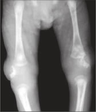
Fig. 23.12: Osteochondroma.
Some important non-malignant bony masses are discussed here. For malignant tumors, see Ch 20.7.
Osteochondroma (Solitary exostosis) is the commonest tumor-like bony swelling in early adolescence, arising from metaphyseal ends of long bones, specially near knee and elbow. Pathologically, it denotes transverse overgrowth (hamartoma) of cartilaginous cells from epiphyseal plate.
Apart from hard bony swelling, these cases may develop local pain, restricted movements, pathological fractures and adjoining nerve/vessel compression. Malignancy is rare. X-ray reveals continuity between cortex and medulla of exostosis and main bone.
Small asymptomatic lesions need no treatment while larger ones may be excised (Fig. 23.12).Enchondroma are benign cartilaginous overgrowths from short long bones, e.g. metacarpals and phalanges. Although usually asymptomatic, some lesions may develop pathological fractures. Ollier disease is a developmental disorder with generalized enchondromatosis.
Osteoid osteoma is a reactive bone lesion, mostly involving cortical areas of femur, tibia and vertebrae. Clinically, these lesions are usually seen beyond 10 years, more common in males and presents with localized pain and tumor-like mass. vertebral lesions may cause spinal cord/root compression. Radiologically, these lesions appear as cystic lesions with an expanding osteolytic lesion and thin cortex of sclerosis. Treatment includes excision/curettage of lesion.
Solitary bone cysts (Unicameral bone cysts) are true cystic lesions at the ends of long bones, usually involving proximal ends of humerus and femur. Clinically, these lesions may be asymptomatic or present with pathological fractures. Diagnosis is based on X-ray and aspiration of the cyst aspiration which may be hemorrhagic. Small cysts heal themselves, while larger ones need surgical curettage and bone grafting. Intralesional methylprednisolone acetate injection is known to heal many lesions.
Aneurysmal bone cyst is solitary rapidly progressive cystic lesion of long bones and vertebrae, probably representing an arteriovenous malformation after trauma or neoplasia. These lesions are rare in childhood, usually presenting as painful swellings in young adults. Along with X-rays, local biopsy is diagnostic, revealing honeycomb of blood-filled cavities. Excision/ curettage with grafting is curative though recurrence is common.
Ganglion is a synovial fluid-filled cyst around the wrist, usually on the dorsal aspect. It denotes a defects in the joint capsule, which allows herniation of the synovium to cause rupture and fluid leakage in soft tissues, followed by reactive fibrosis to wall-off the leak.
In children, most ganglions disappear spontaneously over time and aspiration is indicated only if it is painful, sufficiently large or interferes with normal tendon function. Surgical excision of lesion and tract to the joint space is curative.
Popliteal (Baker) cyst is commonly seen in midchildhood due to distension of gastrocnemius and semimembranosus bursa along the postero-medial aspect of knee by synovial fluid from tendon sheaths. Knee X-ray is normal and diagnosis may be confirmed on USG or aspiration. Most cysts resolve spontaneously after many years and surgery is very rarely indicated.
23.8