MEGALOBLASTIC ANEMIA
Megaloblastic anemia is a hematological term to describe anemia with presence of characteristic megaloblasts in peripheral smear, i.e. large RBCs with finely dispersed nuclear chromatin and disproportionate maturation of nucleus and cytoplasm (Fig.
19.5).It is commonly caused by deficiency of two essential co-factors for synthesis of nucleic acids-vitamin B12 and folic acid. Rare causes include thiamine deficiency/ dependency states and inborn errors of nucleic acid metabolism, e.g. Orotic aciduria and Lesch-Nyhan syndrome.
Peripheral smear in megaloblastic anemia, irrespective of the etiology, characteristically shows:
• Macrocytic RBCs (MCVgt;110 fl) with immature erythroid cells;
• Presence of megaloblasts
• Hypersegmented polymorphs with neutropenia,
• Thrombocytopenia of variable severity
Folic acid deficiency: Normal folic acid requirement in children is ~100 #956;g#8725;day; supplied adequately as folates in most foods and milk (except goat/camel milk). It is absorbed throughout the intestines (Ch 6.3).
Etiology: Folic acid deficiency rarely dietary, usually seen in cases with:
• Poor absorption: Malabsorption syndrome, specially Fish-tapeworm (D. latum) infestations.
• Increased requirements, e.g. hemolytic anemia.
• Impaired folate metabolism due to drugs, e.g. methotrexate, pyrimethamine, and phenytoin.
• Dietary deficiency in babies fed on goat, camel or unfortified powder milk.
Clinically, most cases present at 4-7 months of age with:
• Progressively developing chronic anemia.
• Failure to thrive and chronic diarrhea.
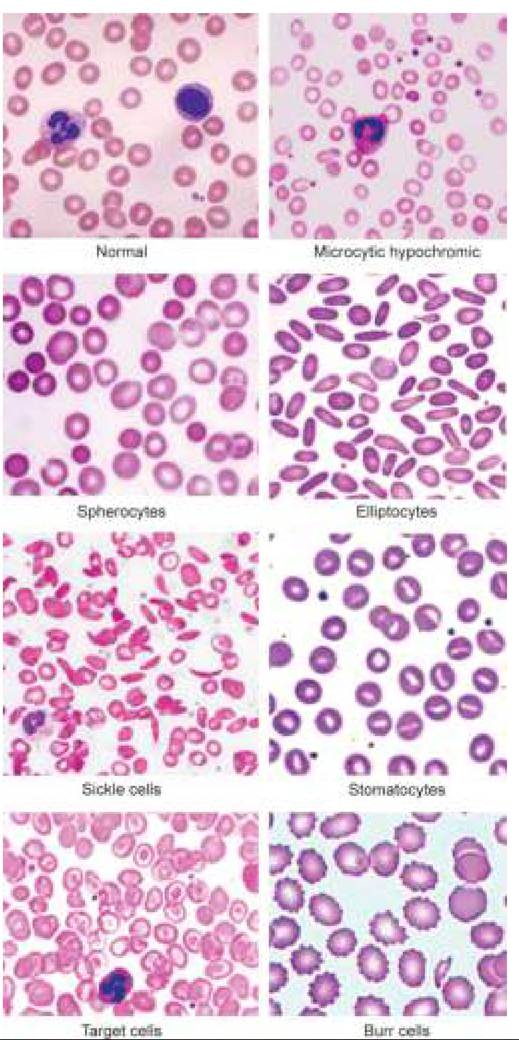
Fig. 19.5: Megaloblastic anemia (peripheral smear).
• Neurological features, e.g. tremors and developmental regression.
• Hyperpigmentation over dorsum of fingers/thigh.
• Bleeding in severe cases due to thrombocytopenia.
A combination of megaloblastic anemia, neurological signs and cutaneous hyperpigmentation suggests possibility of infantile tremor syndrome (Ch 18.7).
Diagnosis is established by:
• Megaloblastic peripheral smear,
• Low serum folate levels lt;3 ng/ml (N: 5-20 ng/ml),
• FIGLU test, i.e. excretion offormiminoglutamic acid in urine after loading with histamine.
Management involves oral or parenteral folic acid therapy (1-5 mg/day for 3-4 weeks), along with vitamin B12 supplements (1000 #956;g#8725;day). Isolated folic acid therapy without B12, may worsen neurological problems associated with co-existing vitamin B12 deficiency despite correction of anemia.
Vitamin B12 deficiency: Daily requirement of vitamin B12 is very low (~1-5 mg), which can be easily fulfilled by dietary cobalamin - vitamin B12 precursor, present in almost all foods. Vitamin B12 absorption requires its hydrolysis by gastric acid and combination with a specific protein — intrinsic factor of Castle (IFc) present in stomach. It is mainly absorbed in distal ileum and transported in plasma as bounded form with a protein- transcobalamin.
Etiology: Dietary deficiency is extremely rare except in total vegans and important causes of Vitamin B12 deficiency in children include:
• Congenital IFc deficiency (juvenile pernicious anemia),
• Malabsorption syndromes involving terminal ileum,
• IFc-B12 receptor deficiency (imerslund syndrome)
• Congenital transcobalamin deficiency
Achlorhydria and presence of Ifc antibodies are other important causes of vitamin B12 deficiency in adults.
Clinically, juvenile pernicious anemia-a rare autosomal recessive disorder of Ifc deficiency, is the prototype of vitamin B12 deficiency, which usually presents at 9-12 months of age, with a triad of:
• Progressively severe anemia,
• Glossitis, i.e.
red smooth and painful tongue, and• CNS signs, e.g. ataxia, paresthesia, hypo/hyperreflexia.
Vitamin B12 deficiency due to other causes presents similarly, except differences in age of presentation and severity.
Diagnosis is established by:
• Megaloblastic peripheral smear,
• Low S. vitamin B12 levels lt; 100 pg#8725;ml,
• Urinary excretion of methylmalonic acid (gt;3.5 mg#8725;d)
• Schilling test.
Schilling test is used to establish presence of vitamin B12 deficiency (Part I) as well as to differentiate between IFc deficiency and malabsorption defects (Part II).
In the first part of test, a small amount of radiotagged (57CO) vitamin B12 is given orally, followed by a flushing dose of 1 mg parenteral vitamin B12 after 2 hours, which leads to excretion of gt;10-30% of radio-tagged dose in urine. Excretion of lt;2% of radio-tagged vitamin B12 indicates its retention in body, due to vitamin B12 deficiency.
In the second part, test is repeated with 30 mg of IFc, given along with the radio-tagged dose. Correction of test results indicates IFc deficiency, while persistent abnormality suggests malabsorption.
Management depends on severity of disease, and generally includes parenteral vitamin B12 (IV#8725;IM) 250-1000 #956;g/day for 2 weeks, then weekly for next 2-3 months, and then monthly doses till correction of anemia. Lower range of dose (0.2 mg) may be used in infants and young children.
Life-long therapy is required in juvenile pernicious anemia or malabsorption disorders. Children with neurological signs must receive parenteral vitamin B12 1000 #956;g (1 mg) daily for 2 weeks followed by alternate week for 6 months and then monthly for life.
Prevention with prophylactic vitamin B12 is advised in cases with malabsorptive states, e.g. autoimmune gastritis or ileal resection.
19.5