QSTEQARTiCULAR TUBERCULOSIS
Skeletal tuberculosis is a late lesion in natural history of tuberculosis, uncommon before 4-5 years of age.
Infection is usually acquired via hematogenous route (disseminated tuberculosis) from pulmonary focus and remains dormant for many years before manifestations.
Trauma, even trivial, is an important preceding factor to facilitate localization of bone infection or progression to disease, due to local vascular injury and inflammation.Pathologically, these lesions are characterized by caseating necrosis, granuloma, bone destruction and ultimately, cold-abscess formation that may trickle to neighboring tissues or through skin (sinus formation).
Common sites of skeletal tuberculosis are: (a) spine (Pott's spine), (b) short bones of hands/feet (tubercular dactylitis) and (c) large joints of lower limbs, e.g. hip.
Pott's spine is the commonest osteoarticular tuberculosis, which primarily involves thoracolumbar spine due to its excessive motility, curvature and maximum stress/strain injury. Apart from hematogenous route, infection may also reach the spine by lymphatics from paravertebral nodes.
Pathogenesis: Commonest site for initial localization of infection in Pott's spine is: (a) metaphyseal region
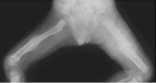
(Courtesy: Dr S Shrivastava)
of vertebrae, adjoining intervertebral disc, followed by (b) central part of body, (c) anterior surface under the anterior longitudinal ligament or rarely, (d) from posterior elements, e.g. lamina or spinous processes. Local lesion may remain dormant or progresses into cold abscess, vertebral destruction and collapse.
Clinically, these cases may present with: (a) localized spinal tenderness or pain during movements, (b) spinal deformity, i.e. kyphosis due to vertebral collapse, (c) signs of compressive myelopathy (20-30%), and/or (d) cold abscess, which may travel along the tissue planes to manifest at distant sites, e.g.
psoas abscess, chest-wall abscess, etc. Constitutional symptoms may be present.Diagnosis must be suspected on clinical grounds and radiological signs, e.g. (a) reduced intervertebral disc spaces, (b) destruction/collapse of vertebrae, and (c) paravertebral shadows of cold abscesses (Fig. 23.11) apart from collaborative evidences for tubercular etiology, e.g. positive chest X-ray, tuberculin test and raised ESR. However, X-rays are unreliable in early disease and CT / MRI is necessary in most cases for confirmation as well as to assess the severity of cord compression.
CT guided biopsy should be attempted in all cases for etiological confirmation for histopathology as well as detection of AFB on staining, culture or CBNAAT, as the gold standard.
Treatment of Pott's spine includes:
• Specific antitubercular therapy including 2HRZE + 10HRZ in drug-sensitive cases, to be modified in resistant cases (Ch 10.13).
• Spinal immobilization in POP jacket/brace during initial 2-3 months, in cases with spinal deformity.
• Drainage of cold abscess, if large or compressing the vital organs.
• General supportive and symptomatic therapy.
Surgery may be indicated in cases with—(a) progressive neurological deficit, (b) paraplegia of recent onset or severe paraplegia, (c) persistent pain with spinal instability, (d) severe or progressive spinal deformity.
Tuberculosis of hip is commonest form of tubercular arthritis, followed by knee involvement, due to weightbearing nature of these joints. Acquired via hematogenous route, infection usually spreads from local osteomyelitis of acetabular roof, femoral head, neck of femur or greater trochanter.
Clinically, these children present with: (a) restricted movements, (b) joint pain on sudden movements (night cry), and (c) limb-length discrepancy due to protective muscular spasm (apparent shortening), joint effusion (lengthening) or articular destruction (true shortening). Acetabular destruction may lead to upward displacement of femoral head (wandering acetabulum).
Cold abscesses from hip usually form in inguinal region. Diagnosis rests on radiological signs: (a) reduced or increased joint space, (b) destruction of articular cartilages, and (c) surrounding osteoporosis. USG or MRI is preferable for early diagnosis and to delineate extent of joint damage. Diagnosis must be confirmed by joint aspiration or synovial biopsy for microbiological confirmation including drug sensitivity profile as well as exclusion of septic arthritis or transient synovitis of hip. Treatment: Apart from specific antitubercular therapy (Ch 10.13), orthopedic management includes traction immobilization for 4-6 weeks, followed by gradual mobilization to retain hip function.In severe cases with cartilaginous destruction, normal mobilization is almost impossible and orthopaedic management aims to achieve a fixed joint in functionally optimal position by prolonged traction/POP immobilization for 4-6 months (Ankylosis), or surgical fusion of joint (Arthrodesis).
Overall functional prognosis is poor despite best treatment, unless hip transplant is possible.
Tubercular dactylitis presents as painful/tender, spindle-shaped swelling of affected phalynx (spina ventosa) with/without discharging sinus. X-ray of affected finger/toe reveals a lytic lesion with minimal sclerosis or new bone formation. Apart from specific antitubercular therapy, immobilization of finger for 4-6 weeks may be helpful. Surgical curettage with bone grafting is necessary in large lesions.
23.6