Brachyspira infections in birds
DESIREE S. JANSSON
Department of Animal Health and Antimicrobial Strategies, National Veterinary Institute & Department of Biomedical Sciences and Veterinary Public Health, Swedish University of Agricultural Sciences, Uppsala, Sweden
The first account of presumed intestinal spirochaetes in birds was published in 1910 and involved morphological descriptions of microorganisms from free-living red grouse (Lagopus lagopus scoticus)(52).
Today, five validated or proposed spirochaetal species assigned to the genus Brachy- spira have been reported to cause enteric disease and production losses in pigs and/or poultry.AETIOLOGY
Brachyspira organisms (trivial name brachyspires) are small (0.2—0.45 ? 1.7—11 μm), helically coiled, highly motile, anaerobic but oxygen- tolerant spirochaetes that colonize the large intestines of some mammalian and avian species (Figure 37.3). They may also be isolated from faecally contaminated environments such as duck ponds. Ultrastruc- turally, brachyspires exhibit typical spirochaetal features, including subterminally attached, bipolar periplasmic flagella that reside in the space between a loosely attached lipid bilayered outer membrane and the protoplasmic cell cylinder. A wave-like or rotational motility centred around the cell axis is an important, albeit not universally present, spirochaetal feature. The spirochaetal design allows brachy- spires to penetrate and move efficiently in intestinal mucin,
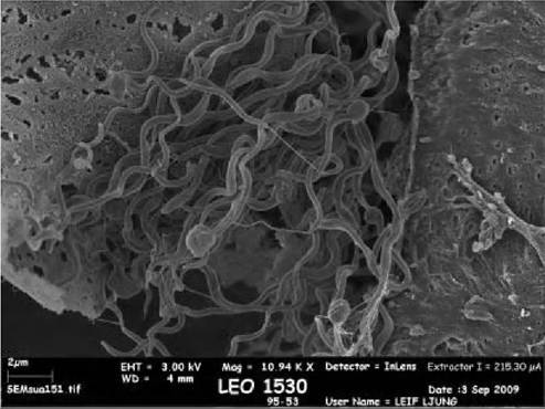
FIGURE 37.3 Scanning electron micrograph of ‘B. suanatina cells. Image: DS Jansson and L Ljung.
which immobilizes many externally flagellated bacteria. Complete genome sequences of B. hyodysenteriae, B. pilosi- coli and B. murdochii were recently published1-53-55).
Genus Brachyspira is the sole member of the family Brachy- spiraceae, which forms a distinct line of descent among the deeply branching genera of the monophyletic phylum Spi- rochaetes. Brachyspires have undergone repeated taxonomic amendments over the last 25 years, and they have previously been classified as belonging to genera Vibrio, Treponema, Serpula and Serpulina. Currently, the genus Brachyspira includes seven validated species, and nine species have been provisionally proposed (Table 37.3).The diseases caused by brachyspires are:
• swine dysentery (pigs; B. hyodysenteriae)
• colonic spirochaetosis, synonyms porcine intestinal spi- rochaetosis, spirochaetal diarrhoea (pigs; B. pilosicoli)
• human intestinal spirochaetosis (B. pilosicoli, B. aalborgι)
• avian intestinal spirochaetosis (B. alvinipull(, B. intermedia, B.pilosicoli in chickens, B. hyodysenteriae in common rhea (Rlhea americana)).
Brachyspira hyodysenteriae is the aetiologic agent of severe necrotizing typhlocolitis in pigs and common rheas, whereas B. pilosicoli (pigs and chickens) and B. intermedia and B. alvinipulli (chickens) cause non-specific colitis and/ or typhlitis, diarrhoea and production losses. In other
TABLE 37.3 List of species assigned to genus Brachyspira.
| Brachyspira speciesa | Published host range |
| B. aalborgi | Human, non-human primates |
| B. hyodysenteriae | Pig, rat, mouse, common rhea (Rhea spp.), mallard (A nas platyrhynchos), chicken, goose |
| B. innocens | Pig, dog, horse, chicken |
| B. pilosicoli | Pig, dog, horse, non-human primates, human, chicken, pheasant, grey partridge, feral waterbirds, common rhea |
| B. intermedia | Pig, chicken |
| B. murdochii | Pig, rat, chicken |
| B. alvinipulli | Chicken, domestic goose, red-breasted merganser (Mergus serrato^, dog |
| iB. canis | Dog |
| iB. pulli | Chicken, dog |
| iB. ibaraki | Human |
| iB. christiani’ | Human |
| iB. suanatina | Pig, mallard |
| iB. corvi | Jackdaw (Corvus monedula), hooded crow ( C. corone cornix), rook ( C. Jrugilegui) |
| cB. rattus' cB. muridarium, iB. muris | Rats and/or mice |
aSpecies within quotation marks have not been fully validated and recognized
hosts, including free-living wild birds and humans, a disease association has not been unequivocally proven.
EPIDEMIOLOGY
Few published data are available regarding geographical distribution, species distribution and prevalence of brachy- spires in free-living wild birds. In Sweden an overall prevalence of 78% from two free- living wild mallard (Anas platyrhynchos) populations was reported1-56). Some isolates were identified as B. hyodysenteriae, whereas others were identified as the closely related proposed species ‘ B. suana- tina,(57), or as B. intermedia, B. pilosicoli, ‘B. pulli, B. alvin- ipulli and closely related genotypes (D.S. Jansson, unpublished observations). More recently, also in Sweden, Brachyspira spp. were isolated from healthy passerine birds of the genera Corvus(58) and Turdus, and from healthy grui- form and charadriiform birds, i.e. Eurasian Coot (Fulica atra) in Sweden and a snowy sheathbill in Antarctica (Chionisalba) (D.S.
Jansson, unpublished observations). In addition to mallards, brachyspires colonize the intestinal tract of a wide variety of other anseriform birds including mute swan (Cygnus olor), whooper swan (Cygnus cygnus), greylag goose (Anser anser), barnacle goose (Branta leucopsis), Eurasian widgeon (A nas penelope), common eider (Somateria mollissima), long-tailed duck (Clangula hyema- lis) and red-breasted merganser (Mergus serrator) (D.S. Jansson, unpublished observations). Parameters such as habitat, diet and physiology (e.g. body temperature, age, stress levels) may possibly explain differences in Brachyspira spp. prevalence between avian species and populations.Because available epidemiological data are limited they do not allow any general conclusions on the possible involvement of free-living wild birds as sources of brachy- spires for domestic animals and humans. Rodents, flies and cockroaches, free-living wild birds and water are considered as possible sources of brachyspires on pig and chickens farms. Swine dysentery and colonic spirochaeto- sis, which are diseases of global importance to the pig industry, have not been reported from European wild boar. Molecular data suggest that zoonotic transfer of B. pilosicoli may occur(59). Brachyspires may remain viable in the environment for days to months if protected by faeces, soil or water. Transmission occurs primarily by the faecal-oral route, but cloacal transmission from water or faecally contaminated surfaces cannot be excluded in gallinaceous and anseriform birds.
PATHOGENESIS AND PATHOLOGY
Virulence attributes of brachyspires and mechanisms of pathogenesis are poorly understood. Proposed virulence factors of B. hyodysenteriae include motility, chemotaxis, oxygen tolerance, lipooligosaccarides, proteases and haemolysins. Brachyspira pilosicoli, and B. aalborg( attach in large numbers by one end to the intestinal epithelium, and this dense bacterial layer speculatively interferes with fluid and nutrient absorption.
In chickens, colonization by brachyspires may be grossly and microscopically inapparent, or is associated with abnormal caecal contents (yellow and frothy) and non-specific typhlitis. Histologically, brachyspires are randomly dispersed in crypts and on epithelial surfaces (B. alvinipull(, B. intermedia, B. pilosicoli), or the bacteria may adhere to the epithelium as described above (B. pilosicoli). Invasive mucosal growth by brachy- spires may occur. In common rheas, severe fibrinonecrotic typhlocolitis has been described (B. hyodysenteriae). Information on gross and microscopic appearance of brachy- spira colonization in free-living wild birds is scarce. Brachyspira hyodysenteriae and ‘B. suanatina, induced epithelial cell changes in mallard caecae by a challenge model, but the birds remained healthy during the trial1-60).CLINICAL SIGNS
Colonization by B. intermedia, B. pilosicoli and B. alvinip- ulli in laying hens produces diarrhoea, reduced egg production and faecally contaminated egg shells1-61), and infection of juvenile common rheas by B. hyodysenteriae causes Abrinonecrotic typhlocolitis and high mortality1-62). Clinical disease in Brachyspira-colonized free-living wild birds is yet to be reported. Interestingly, the recently proposed species ‘B. suanatind, which naturally colonizes pigs and free-living wild mallards, was shown experimentally to induce diarrhoea in pigs, independent of species of origin of the inoculum)57).
DIAGNOSIS
Because of their small size and failure to stain with the Gram stain and routinely applied cytological and histological stains, brachyspires are barely visible by light microscopy. Hence, clear visualization is dependent on dark-field or phase-contrast microscopy, and Warthin-Starry silver staining should be used for histology.
Highly nutritious selective, solid agar media containing antimicrobials, (e.g. spectinomycin, colistin and vancomycin) and 5-10% blood, and anaerobic incubation for 3-9 days at 37-43°C are applied for isolation.
Isolates grow as swarming colonies of varying size and appearance. Species identification may be difficult. Application of phenotypic tests allows presumptive identification, however, more species may colonize avian hosts compared with pigs, and the presence of more than one genotype simultaneously in the same bird is frequently observed. Also, most available diagnostic PCRs tend to lack in sensitivity and/or specificity when applied to avian isolates. Thus, a combination of phenotyping, molecular tests and gene sequencing is often required for definitive identification, and even so, isolates that cannot be assigned to any presently recognized species are occasionally found.SIGNIFICANCE AND IMPLICATIONS FOR ANIMAL HEALTH
Molecular data are accumulating in support of free-living birds, especially anseriforms, as important reservoirs of Brachyspira spp., including those that are pathogenic to domestic animals, and B. pilosicoli, which is potentially pathogenic to humans. The clinical significance in free- living wild bird populations remains to be determined.