Cases
CASE 1
Signalment/history
A 6-year-old, spayed female dog presented for a mucoid, bloody, vaginal discharge. An exploratory laparotomy was performed to remove potential ovarian remnants.
The ovarian pedicles appeared grossly unremarkable. The tissue was excised and submitted for histopathology, and a smear of the vaginal discharge was submitted for cytology (Figures 2.48, 2.49). 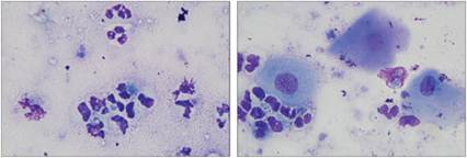
Figures 2.48, 2.49 Bacterial vaginitis. Vaginal smear from a 6-year-old, spayed female Labrador Retriever. Several degenerate neutrophils are present. Many neutrophils contain phagocytized bacterial cocci and rare bacterial rods. Additionally, three squamous epithelial cells are present in 2.49 (Wright–Giemsa, 1,000? magnification).
Cytologic description
The sample was highly cellular. The majority of cells were degenerate neutrophils, most of which were filled with large numbers of bacterial cocci and fewer rods. Scattered mature squamous epithelial cells with abundant rounded to polygonal lightly basophilic cytoplasm and a small round nucleus were present.
Interpretation: septic suppurative inflammation.
Comment
Cornified epithelial cells are typically observed during proestrus and estrus; however, these cells may also come from the cervix or the labia. Given the large numbers of neutrophils and clear indication of infection, this sample is most consistent with bacterial vaginitis. Culture and sensitivity are recommended.
Discussion
It is important to be aware of the normal cytologic appearance of tissues in the area that was sampled before interpreting the data. In this case, low numbers of neutrophils and squamous epithelial cells are a normal finding in vaginal swabs, depending on when the sample was taken during the estrous cycle.
However, squamous epithelial cells can come from several structures near the vagina and should be interpreted with caution in spayed females. The high numbers of neutrophils and intracellular organisms present supported a diagnosis of vaginitis rather than proestrus. Indeed, the histologic sections did not contain any ovarian tissue.CASE 2
Signalment/history
A 10-year-old, intact female Husky presented for a subcutaneous mandibular mass. Six months prior, the patient had been diagnosed and treated for lymphoma. Fine needle aspirates of the right mandibular lymph node (Figure 2.50) and the mass (Figure 2.51) were submitted for cytology.
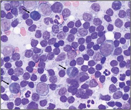
Figure 2.50 FNA of an enlarged right mandibular lymph node from a 10-year-old, intact female Husky. The majority of cells are small lymphocytes. There are moderate numbers of intermediate-sized and large lymphocytes. Increased numbers of plasma cells (arrows) are present (Wright–Giemsa, 1,000? magnification).
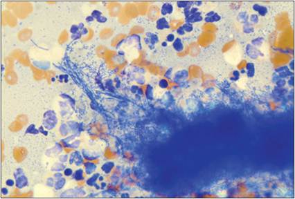
Figure 2.51 FNA from a subcutaneous mandibular mass on a 10-year-old, intact female Husky. Severely degenerate neutrophils are present in large numbers. Several bacterial organisms, including mats of filamentous bacterial organisms, are shown (Wright–Giemsa, 1,000? magnification).
Cytologic description
The lymph node aspirate contained moderate numbers of cells. Most cells were small to intermediate-sized lymphocytes. Few large lymphocytes were seen. There were occasional plasma cells, nondegenerate neutrophils, and eosinophils. The sample from the mass was highly cellular with a small amount of blood in the background. Large numbers of degenerate and nondegenerate neutrophils were present. Lower numbers of macrophages were seen. There were bacterial cocci and rods throughout the slide and within degenerate neutrophils.
Large aggregates of filamentous bacteria surrounded by dying inflammatory cells were noted.Interpretation: lymph node: lymphoid hyperplasia; mass: suppurative inflammation with mixed bacterial sepsis including filamentous bacteria.
Comment
Aerobic and anaerobic bacterial cultures of samples from the mass are recommended.
Discussion
Infection with filamentous bacteria is often associated with pyogranulomatous inflammation. In this case, the suppurative nature of the mass lesion suggests that this was an acute inflammatory process. It is likely that this lesion was identified quickly because the owners were sensitive to swelling in the mandibular area due to the recent diagnosis of lymphoma.
CASE 3
Signalment/history
A 7-year-old, male, neutered domestic short haired cat presented right rear leg swelling and alopecia that had persisted for 4 months. The swelling was firm and erythematous. Bacterial cultures were negative. An FNA of the lesion was performed (Figure 2.52).
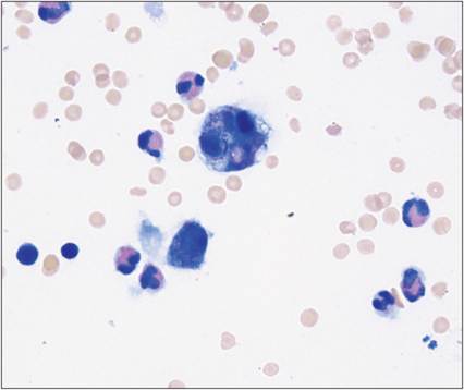
Figure 2.52 FNA of a swollen area on the right rear leg. Several eosinophils with segmented nuclei and pink cytoplasmic granules are shown. There are two macrophages near the center of the image. One macrophage contains phagocytized cellular debris. Two lymphocytes are present at the lower left of the image. A neutrophil is present at the lower right. There are scattered erythrocytes in the background (Wright–Giemsa, 1,000 ? magnification).
Cytologic description
The mass aspirate was moderately to highly cellular with a small amount of peripheral blood. Most cells were eosinophils. Fewer macrophages were seen. Macrophages were arranged individually and in small aggregates. Several macrophages contained cytoplasmic vacuoles and/or phagocytized cellular debris. Occasional neutrophils, small lymphocytes, and intermediate-sized lymphocytes were observed. Rare plasma cells, basophils, and mast cells were noted. No infectious organisms or overtly neoplastic cells were seen.
Interpretation: mass: marked eosinophilic inflammation.
Comment
The clinical appearance of the mass along with the cytologic finding of eosinophilic inflammation supports a diagnosis of an eosinophilic granuloma complex. Biopsy with histopathology is recommended if clinically warranted.
Discussion
Eosinophilic granuloma complex lesions are often associated with underlying allergic and hypersensitivity reactions. Other diseases can cause similar lesions, including ringworm and mite infestation. Further evaluation of cats diagnosed with eosinophilic granuloma complex is critical for proper treatment of the lesions.