Cases
CASE 1
Signalment/history
A mixed-breed dog presented with ocular discharge and marked reddening of the conjunctiva.
Diagnostics
Fluoroscein staining of the right eye revealed marked corneal ulceration and pigmentation (Figure 17.26).
A corneal swab was performed. 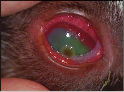
Figure 17.26 Fluorescein-stained cornea from a mixed-breed dog. Note the large pigmented ulcer.

Figure 17.27 Same dog. A mat of fugal hyphae is observed, with parallel walls, septation, and branching arranged in a background of neutrophils and mucocellular debris (Wright–Giemsa, 1,000? magnification).
Cytology
The sample is markedly cellular and consists of a mixed inflammatory population predominated by degenerate neutrophils with few macrophages and small lymphocytes. Thick mats of fungal hyphae are observed with parallel walls, obvious septation, and perpendicular branching (Figure 17.27), consistent with fungal keratitis. Culture revealed Aspergillus flavus.
CASE 2
Signalment/history
A domestic shorthaired cat presented with an eyelid mass.
Diagnostics
Fine needle aspiration of the mass was performed.
Cytology
The sample is highly cellular and consists of many round to polygonal cells arranged individually and in clusters (Figure 17.28). Cells often contain fine to coarse black granular pigment, consistent with melanin. Anisocytosis and anisokaryosis are moderate and some cells contain macronucleoli. These findings are supportive of melanoma and a malignant lesion was suspected due to the degree of atypia. Excision with histopathology was recommended.
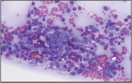
Figure 17.28 FNA of an eyelid mass from a cat. Many pleomorphic melanocytes containing fine to coarse black melanin pigment are observed (Wright–Giemsa, 500? magnification).