Digestive system overview
All animals need a supply of nutrients and oxygen that are obtained via the digestive system and respiratory system, respectively. The digestive system consists of the digestive tract, also called the gastrointestinal or alimentary tract, and its accessory organs.
The accessory organs include the teeth, tongue, salivary glands, liver, pancreas, and gallbladder.The digestive tract is a muscular tube running through the body extending from the mouth to the anus. Contents within the digestive tract are considered outside the body, so in addition to digesting and absorbing nutrients, the digestive tract must act as a barrier blocking the entry of pathogenic organisms.
Functions of the digestive tract
The digestive tract has eight functions:
1. Ingestion. This is the active process of bringing material into the oral cavity.
2. Propulsion. Ingested materials are moved through the digestive tract by swallowing and peristalsis (peri = around + stalsis = constriction), which involves alternating waves of contraction and relaxation of muscles along the digestive tract wall (Fig. 17.1), and is the major propulsive mechanism moving food through the tract.
3. Mechanical processing. Material entering the digestive tract is physically reduced in size. This begins in the oral cavity where food is crushed
Anatomy and Physiology of Domestic Animals, Second Edition. R. Michael Akers and D. Michael Denbow. © 2013 John Wiley & Sons, Inc. Published 2013 by John Wiley & Sons, Inc.
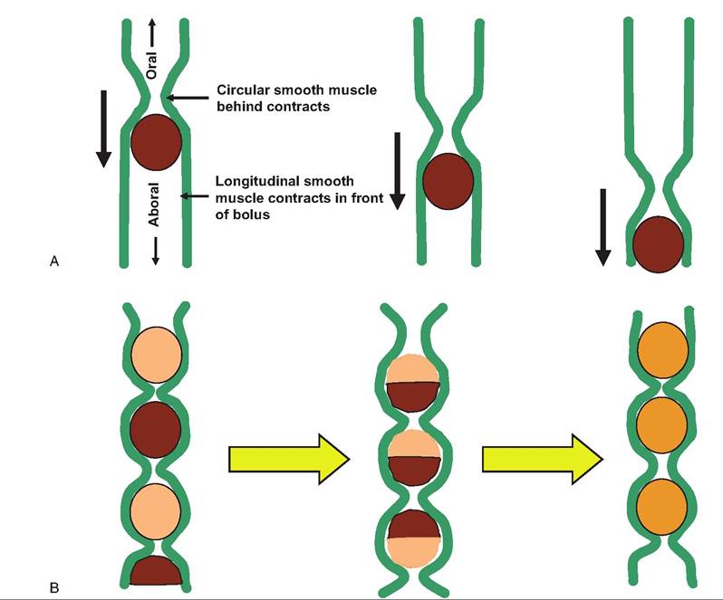
Fig. 17.1. Peristalsis and segmentation. (A) During peristalsis, the circular smooth muscle layer behind the bolus contracts while that in front of the bolus relaxes. Conversely, the longitudinal smooth muscle layer behind the bolus relaxes while that in front of the bolus contracts.
This increases the diameter of the lumen in front of the bolus while constricting the diameter of the lumen behind the bolus. This results in propulsion of the bolus down the digestive tract. (B) During segmentation, nonadjacent sections of the digestive tract contract and relax, resulting in mixing of the contents.and sheared before being propelled along the digestive tract. The reduction in size of ingested material increases its surface area, thereby facilitating enzymatic digestion. In the case of ruminant animals, food materials are also moved from the stomach back to the mouth for further reduction in particle size. Food is also churned along the digestive tract by segmental contractions (Fig. 17.1), which further mix the contents with digestive juices, but do not advance their movement.
4. Digestion. Following reduction in size, ingested nutrients are chemically broken down into particles small enough for absorption. Although simple molecules such as monosaccharides and amino acids can be absorbed without further reduction in size, macromolecules such as protein, DNA, polysaccharides, and triglycerides must first be reduced into smaller molecules. Specific enzymes complete such reduction.
5. Secretion. Water, mucus, acids, enzymes, buffers, and salts are released into the lumen of the digestive tract along its length. Secretions come from epithelial cells and glandular organs.
6. Absorption. Along the length of the digestive tract, nutrients including organic substrates, electrolytes, vitamins, and water pass from the lumen into the body. In addition to absorbing ingested nutrients, the digestive tract must absorb secreted water, salts, and other secreted material. Failure of such absorption will result in dehydration.
7. Excretion. The digestive tract is a site of elimination of waste products. Such waste products can be eliminated via either defecation or egestion.
8. Immunity. The digestive tract must provide a substantial barrier to prevent the entry of pathogens into the body.
The digestive tract acts not only as a physical barrier but also has an innate immune system.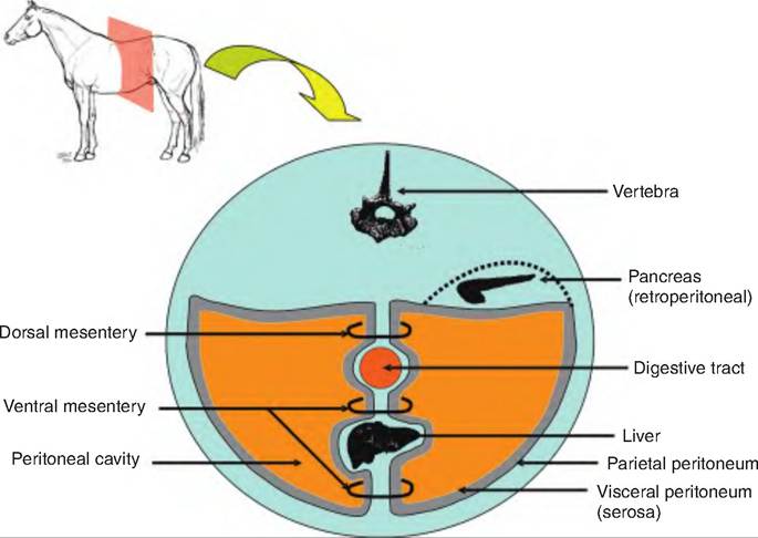
Fig. 17.2. Peritoneum and peritoneal cavity. The cross section through the abdominal cavity of the horse shows the visceral and parietal peritoneum, and the dorsal and ventral mesentery. Although not shown, the peritoneal cavity is filled with organs. Note that some organs, such as the pancreas, are located in a retroperitoneal position, or outside the peritoneal cavity.
Peritoneal cavity
The peritoneal cavity is formed from a serous membrane, called the peritoneum, which lines the abdomi- nopelvic cavity, forming the largest serous membrane in the body (Fig. 17.2). It consists of a layer of simple squamous epithelium, the mesothelium, with an underlying connective tissue layer. The peritoneal membrane has a serosa, or visceral layer, that covers the organs in the peritoneal cavity, and a parietal peritoneum that lines the inner surface of the body wall.
The peritoneal membrane produces peritoneal fluid providing lubrication between the serosa and parietal layers, called the peritoneal cavity, thus reducing friction and irritation. Diseases of the liver, kidney, and heart can cause increases in this fluid production producing an abdominal swelling called ascites. Accumulation of this fluid can distort the internal organs, causing pain and discomfort.
Mesenteries
The mesentery consists of two layers of serous membranes fused back to back, and suspends portions of the digestive tract from the body wall. The mesenteries have three functions: (1) to provide a route for blood vessels, lymphatic vessels, and nerves to travel to the digestive system; (2) to hold organs in place; and (3) to store lipid.
During embryonic development, the digestive organs are suspended from the body wall by the dorsal and ventral mesentery. The ventral mesentery largely disappears except on the ventral surface of the stomach, between the stomach and liver and between the liver and the ventral abdominal wall.
The mesenteries are named for the organs they supply (e.g., mesoduodenum, mesoileum, mesocolon).The omentum refers to those portions of the mesentery connecting the stomach to the abdominal organs or abdominal wall. In animals with simple stomachs, such as carnivores, pigs, and horses, the greater omentum connects the greater curvature of the stomach to the dorsal abdominal wall (Fig. 17.3). It folds over itself forming deep and superficial layers (i.e., four layers). It normally contains considerable adipose tissue. The lesser omentum connects the lesser curvature of the stomach and initial segment of the duodenum with the liver. The falciform ligament attaches the liver to the ventral midline while the hepatoduodenal ligament connects the liver to the proximal duodenum.
In ruminants, the superficial and deep portions of the greater omentum attach to the left side of the rumen and the right side of the rumen, respectively. They tract toward the right side of the animal, attaching to the intestine and then to the right abdominal wall. Although most abdominal organs are located within the peritoneum, some are located between the posterior parietal peritoneum and the posterior
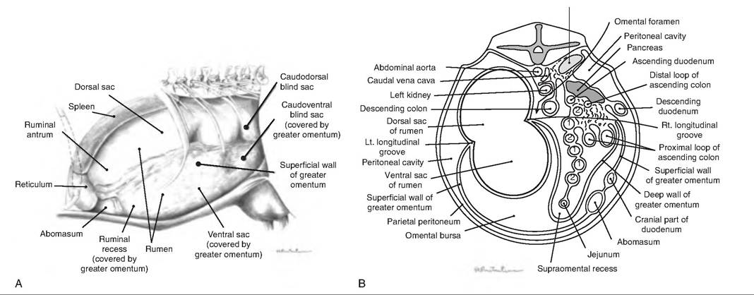
Fig. 17.3. Arrangement of mesenteries in ruminants. (A) In this view of the left side of a large ruminant, the greater omentum is visible. (B) A cross section of the flank of a large ruminant showing greater omentum and peritoneum. (Reprinted from Constantinescu and Constantinescu, 2004. Used by permission of the publisher.)
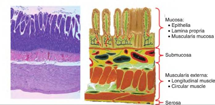
Fig. 17.4. Basic structure of the digestive tract. The four basic layers of the digestive tract, from the lumen outward, are the mucosa, submucosa, muscularis externa, and serosa.
abdominal wall, and are thus outside this cavity and are said to be retroperitoneal (retro = behind). These organs include the kidneys, adrenal glands, ureters, duodenum, ascending colon, descending colon, and pancreas. Those organs whose mesenteries remain inside the peritoneal cavity are called intraperitoneal or peritoneal organs.
Blood supply of the digestive organs
The splanchnic circulation serves the digestive organs and hepatic portal system. The arteries of this system include the hepatic, splenic, and left gastric branches of the celiac trunk serving the spleen, liver, and stomach, respectively, and the mesenteric arteries serving the small and large intestines. At rest, the splanchnic circulation receives approximately 25% of the cardiac output.