Indications
Bone marrow examination has a central role in the diagnosis of hematologic and infectious conditions, and for detecting and staging potentially metastatic neoplasms. Ideally, both marrow aspiration cytology and core (trephine) biopsy should be performed; the former facilitates detailed cell morphology assessment, differential cell counting, and identification of infectious agents, while the latter enables determination of overall cellularity, hemosiderin stores, and tissue architecture.
Any abnormal complete blood cell count (CBC) finding that cannot be explained by extramedullary causes is a primary indication for marrow evaluation. Specific examples of CBC abnormalities that should prompt marrow examination include persistent and unexplained cytopenia (non-regenerative anemia, neutropenia, or thrombocytopenia); dysplasia in one or several cell lines (Figure 19.1); inappropriate or persistent rubricytosis (Figure 19.2); ovalocytosis (Figure 19.3); and the presence of blasts or other atypical cells in circulation (Figures 19.4, 19.5). Assessment of marrow to investigate possible lytic lesions or metastatic neoplasms constitutes a secondary indication. Marrow aspiration and biopsy are relatively invasive procedures and, therefore, indications should be clearly established. Marrow samples should be interpreted in light of the history, physical examination, concurrent CBC findings, and results of ancillary diagnostic assays such as serum biochemistry, infectious disease serology, and imaging. 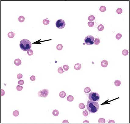
Figure 19.1 Blood smear from a dog with neutropenia, non-regenerative anemia, and neutrophil dysplasia. Note large hyposegmented neutrophils with condensed chromatin (arrows), 600? magnification.
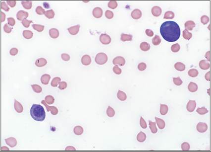
Figure 19.2 Blood smear from a dog with persistent neutropenia (0.5–0.9 ? 109/L), anemia (hematocrit 0.20–0.29 L/L), thrombocytopenia (45–62 ? 109/L), low-grade rubricytosis, and atypical cells, 600? magnification.
Clinical assessment failed to reveal infection or inflammation, and a diagnosis of chronic neutrophilic leukemia was established.
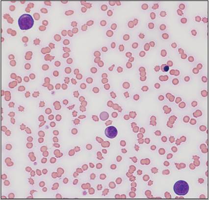
Figure 19.3 Blood smear from a cat with rubricytosis, 400? magnification.
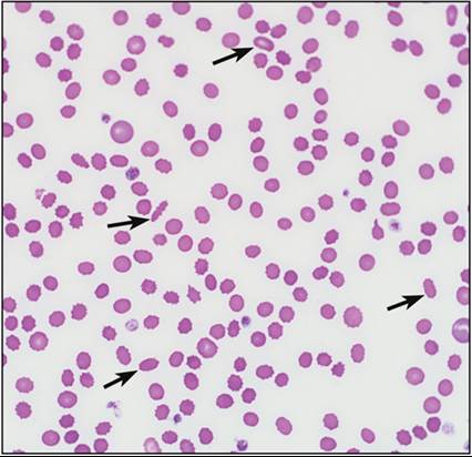
Figure 19.4 Blood smear from a dog with non-regenerative anemia characterized by ovalocytosis and acanthocytosis (arrows), 400? magnification.
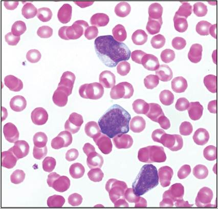
Figure 19.5 Blood smear from a dog with anemia, thrombocytopenia, and blasts, 1,000? magnification.