Inflammation
Inflammation of the eyelid, termed blepharitis, may involve one or both eyelids and can present as focal or diffuse lesions. The inflammatory populations that occur in the eyelid parallel those found in the skin of other parts of the body and follow a similar algorithm of differentials.
Certain diseases may only involve the eyelids, while others, such as demodectic and sarcoptic mange, include the eyelids in addition to other lesions on the face and body (Aroch et al., 2008). Infectious, immune-mediated, and foreign body reactions should be considered if suppurative inflammation is present. Primary bacterial blepharitis in adult dogs is often caused by Staphylococcus spp. and Streptococcus spp., and many degenerate neutrophils are typical on cytologic examination (Stiles, 2012). Juvenile cellulitis (also known as puppy pyoderma or puppy strangles), a presumed immune-mediated condition that causes severe sterile abscessation and edema of the muzzle and mandibular lymph nodes in puppies, often involves the eyelids (Bassett et al., 2005). Cytology of pustules reveals aseptic suppurative to pyogranulomatous inflammation.Eosinophilic inflammation alone or as part of a mixed inflammatory population may result as part of a hypersensitivity response to insect stings or bites. Additionally, immune-mediated eosinophilic plaques may involve the eyelids and reveal marked eosinophilic inflammation with or without lower numbers of other inflammatory cells. Pyogranulomatous inflammation secondary to fungal infections (dermatophytes, Blastomyces dermatitidis) and foreign body reactions may also occur (Figure 17.1). Patients infected with Leishmania infantum often develop eyelid lesions, which can vary in presentation including dry dermatitis and alopecia, blepheredema, ulcers, and discrete granulomas (Pena et al., 2000). Cytology of granulomas reveals numerous macrophages with or without neutrophils, and in many cases Leishmania infantum amastigotes are present (Pena et al., 2000).
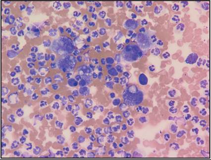
Figure 17.1 FNA of an eyelid mass from a dog. Many vacuolated macrophages and low numbers of multinucleated macrophages are present (Wright–Giemsa, 500? magnification).
Common non-neoplastic masses that occur on the eyelid include the chalazion, hordeolum, and cystic structures. Chalazion is a lipogranuloma of the meibomian gland, and build-up of the secretory product leads to granulomatous inflammation (Figure 17.2; Stades & van der Woerdt, 2013). Many macrophages and lower numbers of multinucleated giant cells and small lymphocytes are most commonly seen on cytology. Hordeolum is focal abscessation of the sebaceous glands of the eyelid, including the meibomian glands and glands of Zeis, and aspiration will reveal suppurative inflammation. Lastly, Meibomian cysts can occur and typically consist of foamy macrophages and cholesterol crystals in highly proteinaceous material on cytologic examination.
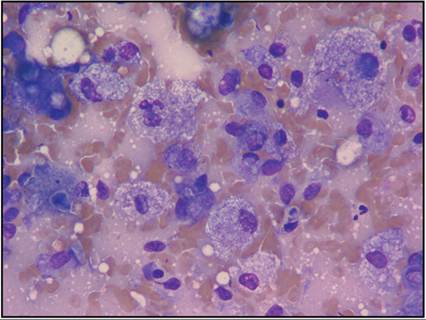
Figure 17.2 FNA of an eyelid mass from a dog. Note the abundance of vacuolated macrophages with low numbers of multinucleated giant cells. No infectious organisms were identified (Wright–Giemsa, 1,000? magnification).
Neoplasia
Neoplastic masses of the eyelid are most often benign (Krehbiel & Langham, 1975). The most common tumor is the meibomian adenoma, with meibomian adenocarcinoma being far less common ( Krehbiel & Langham, 1975). Meibomian adenomas appear cytologically similar to aspiration of any other sebaceous glandular tissue, with many clusters of epithelial cells filled with discrete clear cytoplasmic vacuoles. Melanomas are the second most common tumor of the eyelid and appear cytologically similar to melanomas of other sites but are typically less aggressive. Fibroma, fibrosarcoma, papilloma, squamous cell carcinoma (SCC), mast cell tumor (Figure 17.3), lipoma, granular cell tumor, histiocytoma, and meibomian gland epithelioma (Figures 17.4a, b) are all less common but reported (Krehbiel & Langham, 1975; Lu & Dubielzig, 2012).
Histiocytosis of Bernese Mountain Dogs frequently involves the eyelids, and aspiration of the nodules reveals neoplastic histiocytes with marked criteria of malignancy (Moore, 1984). 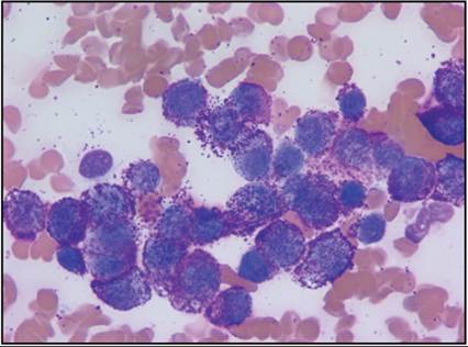
Figure 17.3 FNA of an eyelid mass from a dog. Many mast cells are present, which contain many dark purple cytoplasmic granules (Wright–Giemsa, 1,000? magnification).
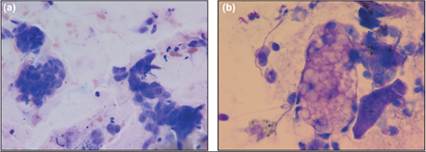
Figures 17.4a,b Additionally, low numbers of epithelial clusters with abundant cytoplasmic clear vacuolation consistent with benign sebaceous epithelial cells are present (Wright–Giemsa: a, 500? magnification; b, 1,000? magnification). Many cohesive clusters of benign epithelial cells with a small amount of basophilic cytoplasm, round nuclei, and indistinct nucleoli are present.
Conjunctiva
The conjunctiva is a mucous membrane that lines the inner surface of the eyelids and nictitating membrane and then reflects back onto the anterior portion of the globe. The palpebral conjunctiva consists of pseudostratified epithelium with goblet cells and lymphoid follicles. The bulbar conjunctiva consists of nonkeratinized squamous epithelium (Figure 17.5; Raskin, 2010). As this tissue approaches the limbus it is common to see melanin granules coating the epithelial cells (Figure 17.6; Young & Prasse, 2007). The melanin granules are easy to confuse with bacteria. Melanin granules have a brownish-gold staining with most Romanowsky stains while bacteria will stain pale to deeply basophilic. Epithelial cells from most tissues are cohesive and can be found in sheets and clusters; however, squamous epithelial cells can be more individualized. Columnar epithelial cells are elongate and on cytology can appear similar to spindle cells. The other types of epithelial cells are more likely to be round with a centrally placed nucleus and variable amounts of cytoplasm depending on the location within the conjunctiva (Figure 17.7).
Goblet cells have voluminous amounts of cytoplasm, which is filled with eosinophilic globular material. Cytologically, lymphoid follicles will consist of a mixed lymphoid population, predominated by small lymphocytes with few large lymphocytes and plasma cells. 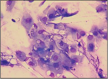
Figure 17.5 Conjunctival scrape from a dog. One cluster of nonkeratinized squamous epithelial cells is present. The cells have abundant amounts of basophilic cytoplasm and a single round nucleus (Wright–Giemsa, 1,000? magnification).
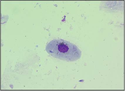
Figure 17.6 Conjunctival swab from a dog. A single squamous epithelial cell coated in melanin granules is present (Wright–Giemsa, 1,000? magnification).
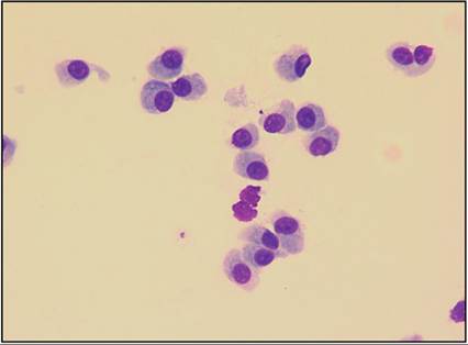
Figure 17.7 Conjunctival scrape from a dog. Scattered columnar epithelial cells are observed. These cells are rectangular to spindle in shape with single round nuclei (Wright–Giemsa, 1,000? magnification).
Cytologic samples of conjunctiva can be obtained by multiple techniques, including swabs, scrapes, and imprints (Bauer and Speiss, 1996). The sampling technique will impact the types of epithelial cells observed. An imprint will likely sample the surface epithelium and result in squamous epithelial cells being the most prominent. A scrape will sample deeper areas of the tissue, so goblet cells and small lymphocytes will also be observed in addition to epithelial cells. The epithelial cells from the deeper layers are more likely to have a centrally placed nucleus and a moderate rim of pale basophilic cytoplasm (Bolzan et al., 2005). Common findings from cytology from healthy conjunctiva include a predominance of epithelial cells with a small amount of mucus and few small lymphocytes (Lavach et al., 1977).
Impression cytology, performed by applying a cellulose filter to the superficial layers of epithelial cells, produces cellular samples while also preserving tissue architecture (Perazzi et al., 2017).Inflammation
Cytologically, inflammation of the conjunctiva is characterized by the type of inflammatory cells seen (e.g. >85% neutrophils is consistent with suppurative inflammation). Nonspecific changes that may accompany inflammation include lymphoid hyperplasia, epithelial hyperplasia, and squamous metaplasia with keratinization of squamous epithelial cells (Wilcock, 2007). Excessive mucus is often produced resulting in thick eosinophilic strands of mucinous material (Figure 17.8). Conjunctivitis can be primary or secondary. Cytologically, unless the causative agent is identified, the underlying cause is often difficult to discern. Neutrophils are commonly associated with bacterial, viral, and fungal infections but can also be seen secondary to keratoconjunctivitis sicca and allergic disease. The condition of the neutrophils should be evaluated. If the neutrophils are undergoing karyolysis or appear degenerate, this can be associated with bacterial infection. Degenerate neutrophils have pale swollen nuclei that are starting to lose the filamentous structure connecting the lobules of the nucleus (Figure 17.9). Fungal conjunctivitis has been reported in cats, including primary conjunctival sporotrichosis (Spinelli et al., 2021) and disseminated histoplasmosis with conjunctival and/or nictitating membrane involvement (Ewald et al., 2020). Small numbers of lymphocytes and plasma cells can be observed in normal conjunctiva but also can be associated with inflammation or immune stimulation. In true lymphoplasmacytic inflammation, the lymphoid inflammatory cell population predominates over the epithelial cells. Immune-mediated diseases, such as pannus, medial canthal blepharitis of Long-haired Dachshunds, and follicular conjunctivitis, will have a very cellular infiltrate predominated by small lymphocytes with many plasma cells (Figure 17.10; Raskin, 2010).
In other immune-mediated diseases, such as pemphigus, neutrophils and eosinophils are the primary inflammatory cells. Allergic conjunctivitis can result in a mixed cellular infiltrate with a high number of eosinophils and scattered mast cells (Figure 17.11). 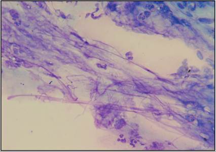
Figure 17.8 Conjunctival swab from a dog. Several thick strands of eosinophilic mucinous material are observed, admixed with scattered neutrophils (Wright–Giemsa, 1,000? magnification).
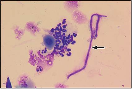
Figure 17.9 Conjunctival scrape from a dog. A group of degenerate neutrophils are surrounding a squamous epithelial cell. The epithelial cell has a ‘fried egg’ appearance with a centrally placed nucleus and a moderate rim of basophilic cytoplasm. A fungal hyphal structure (indicated by an arrow) is also observed (Wright–Giemsa, 500? magnification).
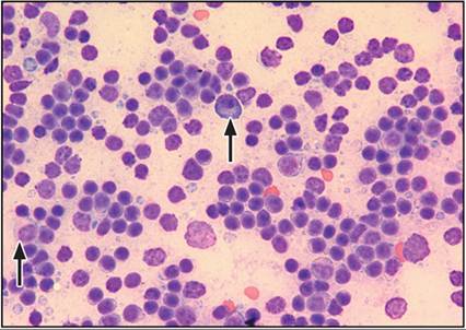
Figure 17.10 Conjunctival scrape from a dog with lymphoplasmacytic conjunctivitis. The sample is cellular, and a mixed lymphoid population predominated by small lymphocytes with few plasma cells (indicated by arrows) is observed. Note the perinuclear clearing or prominent Golgi apparatus in the cytoplasm of the plasma cells (Wright–Giemsa, 500? magnification).
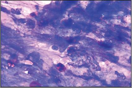
Figure 17.11 Conjunctival swab from a cat with allergic conjunctivitis. The sample is cellular. A mixed inflammatory population, predominantly neutrophils with many eosinophils, is observed embedded in a thick mat of eosinophilic mucinous material (Wright–Giemsa, 1,000? magnification).
Cellular inclusions may be observed in conjunctival specimens. These inclusions can be identified in inflammatory cells and conjunctival epithelial cells and may be intracytoplasmic or intranuclear. Infectious organisms such as Chlamydia spp., Mycoplasma spp., or distemper virus can be found in the cytoplasm of epithelial cells (Figures 17.12a, b). However, more commonly the cytoplasmic inclusions are benign structures such as melanin granules or ophthalmic ointment (Figure 17.13), which are easy to misinterpret as an infectious etiology (Young & Taylor, 2006). Viral inclusions have been reported to occur (Raskin, 2010) with feline herpesvirus and canine distemper, but are infrequently encountered in cytologic specimens (Hillstrom et al., 2012).
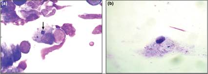
Figures 17.12a,b Conjunctival swab from a cat with an ocular discharge. (a) A large epithelial cell with a chlamydial inclusion (arrow) is identified (Wright–Giemsa, 1,000? magnification). (Courtesy Dr. Richard Meadows.) (b) Conjunctival scrape from a dog with a bilateral ocular discharge, nasal discharge, and a fever. One epithelial cell is observed containing a single basophilic cytoplasmic inclusion consistent with canine distemper virus (Wright–Giemsa, 400? magnification).
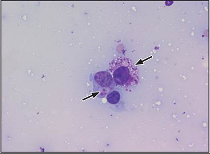
Figure 17.13 Conjunctival swab from a dog. The patient had been receiving ophthalmic ointment in both eyes for 1 week prior to sampling. A single epithelial cell containing several eosinophilic inclusions consistent with ophthalmic ointment is indicated by the arrow on the left. A single mast cell is also present (right arrow) (Wright–Giemsa, 1,000? magnification).
Neoplasia
Conjunctival neoplasia may appear as a discrete mass or an irregular thickening of the conjunctiva. If a mass is identified, fine needle aspiration (FNA) is the most efficient method to obtain a cytologic sample; however, conjunctival scrapes can also be beneficial. Many tumors have been described and include mast cell tumor, lymphoma, melanoma, hemangioma, hemangiosarcoma, and SCC, with SCC as the most common (Pirie et al., 2006; Maggs, 2008; Schobert et al., 2010; Fife et al., 2011; Olbertz et al., 2012). Cytologically, SCCs appear similar regardless of the location. The cells are round to polygonal with varying amounts of pale basophilic to keratinized cytoplasm. The nuclei vary greatly in size with a coarsely stippled to pyknotic chromatin. A ring of perinuclear vacuoles can be observed (Figure 17.14). Occasionally, neutrophils can be seen coating the cytoplasm. It is not uncommon to have a significant suppurative inflammatory response accompanying the tumor. The presence of inflammation makes SCC difficult to differentiate from squamous dysplasia secondary to inflammation.
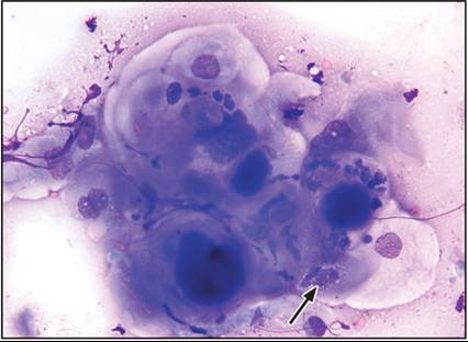
Figure 17.14 Conjunctival aspirate from a conjunctival swelling in a cat with squamous cell carcinoma. A cluster of neoplastic squamous epithelial cells, occasionally coated in neutrophils. The arrow indicates a perinuclear ring of cytoplasmic vacuoles (Wright–Giemsa, 1,000? magnification).
Mast cell tumors (MCTs) can be readily diagnosed with cytology via FNA (Fife et al., 2011). MCTs are round cell tumors and the neoplastic cells are typically individualized and round. Mast cells have a moderate amount of cytoplasm filled with dark purple granules (Figure 17.15). Stains such as Diff-Quik® do not consistently stain the granules, which may decrease the diagnostic yield of in-house cytology.
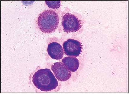
Figure 17.15 Conjunctival scrape from a dog with conjunctival swelling with mast cell neoplasia. Highly granular mast cells are identified (Wright–Giemsa, 1,000? magnification).
Conjunctival melanomas typically consist of very cellular samples often containing high numbers of melanin granules, although amelanotic melanomas have been reported (Schobert et al., 2010). Amelanotic melanoma can be difficult to diagnose cytologically without immunocytochemical or immunohistochemical staining. Melanotic melanomas are relatively easy to diagnose with cytology since the cells are frequently heavily pigmented (Figure 17.16). This abundance of pigment may obscure nuclear detail.
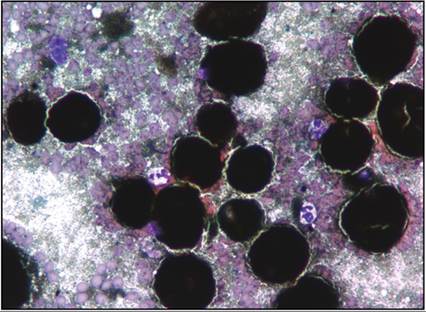
Figure 17.16 Conjunctival aspirate from a mass in a cat with a conjunctival melanoma. The sample is highly cellular with many highly granular neoplastic melanocytes. The cells are so granular, the nuclear detail cannot be visualized. Melanin granules and red blood cells are present in the background (Wright–Giemsa, 1,000? magnification).