Normal gastrointestinal cytology
The alimentary or gastrointestinal tract (GIT) in mammals is broken up into four major sections: the esophagus, the stomach, the small intestine, and the large intestine/colon and rectum.
It is essentially a muscular tube lined by a mucous membrane. The cellular constituents making up the normal architecture of each section are specific and related to the function of each in order to aid in the passage and digestion of food while assisting in protection against ingested pathogens. All sections are comprised of four distinct layers of tissue: the mucosal layer at the luminal surface, a submucosal layer, a layer of smooth muscle, and an outer serosal/adventitial layer. These ‘tunics’ are designed for optimal protection, passage, absorption of water and nutrients, and excretion of waste (Figure 7.1). 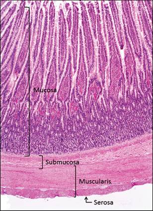
Figure 7.1 Histologic section of the intestine. Four distinct layers make up the intestinal ‘tunics’: mucosa, submucosa, muscularis, and serosa (H&E, 40? magnification) (courtesy Linda Meola).
Tunica mucosa
The epithelial mucosa lines the GI lumen and is divided into three distinct layers (Figure 7.2): the epithelial lining (lamina epithelialis), which is unique in each section of the intestine; a lamina propria layer, which contains lymphoid cells to assist in local immunity (Figure 7.3); and a thin layer of smooth muscle, the muscularis mucosae, which provides for local movement. Aggregates of lymphoid cells are found throughout the length of the GIT making up the ‘gut-associated lymphoid tissue’ (GALT) (Figure 7.4).
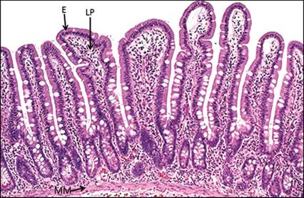
Figure 7.2 Histologic section of gastrointestinal mucosa. The mucosa is comprised of three layers from outer to inner: the epithelial layer (E), the lamina propria (LP), and the muscularis mucosae (MM) (H&E, 200? magnification).
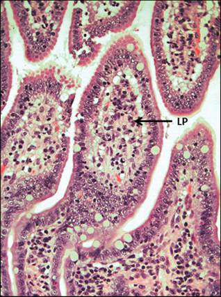
Figure 7.3 Histologic section of small intestinal mucosa, feline. Lymphoid cells comprise the primary immune cell population of the lamina propria (LP, arrow) (H&E, 400? magnification).
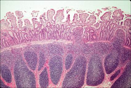
Figure 7.4 Histologic section of the intestinal mucosa, canine. Lymphoid follicles (GALT) in the mucosa of the gastrointestinal tract (H&E, 40? magnification) (courtesy Barry Cooper).
Tunica submucosa
The submucosal layer is comprised of loose collagenous connective tissue, which contains blood and lymphatic vessels and nerves for support of the mucosa.
Tunica muscularis
The muscular tunic is comprised of smooth muscle layers organized perpendicularly to each other for peristaltic activity.
Tunica serosa/adventitia
The outer layer of the alimentary tract consists of loose supporting tissue and adipose with associated vessels and nerves. Within the abdominal cavity, this layer is referred to as the serosal layer and is lined by simple squamous epithelium (mesothelium).