OTHER HERPESVIRUS INFECTIONS
Frederik widen and dolores gavier-widen
National Veterinary Institute (SVA) and Swedish University of Agricultural Science, Uppsala, Sweden
Infection of European hedgehogs (Erinaceus europaeus) with a herpesvirus classified as Erinaceid herpesvirus 1
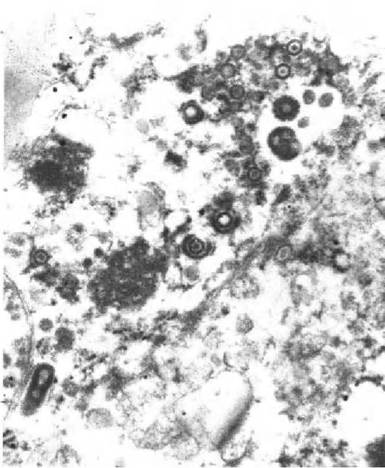
FIGURE 1.5 Electron microscopy of herpesvirus from a hedgehog with hepatitis cultured on primary bovine foetal skin cells.
(ErHV-I) has been described in Europe)88-90). Herpesviruslike particles were demonstrated in the liver of a hedgehog with hepatitis in the UK)88), fatal herpesvirus infection was detected in a 3-month-old hedgehog in Sweden(89), and a severe viral meningoencephalitis caused by herpesvirus in an orphan hedgehog brought to a wildlife rehabilitation centre was described in Switzerland)90). Herpesvirus was isolated on primary bovine fetal skin cells from the liver of the hedgehog in Sweden )Figure 1.5), and a cytopathic effect characteristic of alphaherpesvirus was observed within 48 hours)89). Herpesvirus particles were observed by electron microscopy in the case in the UK)1). Abundant viral antigen was detected by immunohistochemistry in the nucleus and cytoplasm of neurons and glial cells using antibodies for human herpes simplex virus type 1 and 2)90). Little is known about the epidemiology. The case in Britain affected an adult female. In Sweden, a litter of four 2-week- old orphan hedgehogs had been hand reared up to the age of 3 months and placed with adult hedgehogs. Three of the hedgehogs in the litter died within 2 days of mixing the groups, and the remaining one on the fifth day. Only one was submitted for postmortem examination. The case in Switzerland affected a young female. In the cases in the UK and Sweden, the liver was the most severely affected organ, showing coagulative necrosis, fatty degeneration and mild inflammatory infiltrate )Figure 1.6).
Intranuclear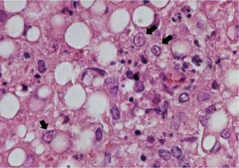
FIGURE 1.6 Histological section of liver of a European hedgehog with fatal herpesviral hepatitis, showing necrosis of hepatocytes and fatty degeneration. Intranuclear acidophilic inclusion bodies are observed in hepatocytes (arrows). Photo: D. Gavier-Widen, SVA.
acidophilic inclusion bodies were observed in hepatocytes (Figure 1.6 ) and Kupffer cells. Similar lesions were observed in the adrenal glands(89). In the case in Switzerland, multifocal perivascular cuffing, diffuse meningeal infiltration with lymphocytes and plasma cells, numerous eosinophilic intranuclear inclusion bodies in neurons and glial cells of the cortex and to a lesser content of the thalamus and the brainstem and neuronal necroses were observed(90). The clinical course in all the cases appeared acute and the hedgehogs were in good bodily condition. The case of meningoencephalitis clinically showed progressive incoordination, circling, and finally loss of appetite. It can be concluded that a potentially fatal alphaherpesvirus infection of hedgehogs occurs in Europe but that the virus is poorly characterized and information regarding the epidemiology and zoonotic aspects is not available. Whether hepatitis and meningoencephalitis is caused by the same herpesvirus genus remains a speculation.
Applying molecular methods, seven gamma- and one betaherpesvirus, belonging to seven different European bat species, were identified in 15 individual bats in Germany(91). As the bats had been found at different locations and on different dates, it was considered unlikely that they originated from the same roost populations. None of the bats showed histological lesions that could be attributed to herpesviral infection. However, it is known from other species that infection with gamma- and betaherpesviruses often has a subclinical course, and the pathological significance/potential of the herpesviruses in these bats could not be elucidated.
A herpesvirus (genus Rhadinovi- rus, subfamily Gammaherpesvirinae) was detected by nested PCR in an adult female serotine bat (Eptesicus serotinus) submitted to a rescue centre in Hungary. The bat had died with signs of icterus and anorexia within a day, in spite of supportive therapy. The causative role of the herpesvirus could not be proven(92).Mustelid herpesvirus 1 (MusHV1; genus Rhadinovirus, subfamily Gammaherpesvirinae) has been reported to be frequent in badgers (Meles meles) in the British Isles. A high percentage offree- ranging badgers sampled at two geographically distinct locations (southwest ofEngland and the Republic ofIreland) were found positive by PCR. The seroprevalence investigated by an in-house indirect ELISA, revealed antibodies in 36 out of 110 badgers tested. The antibody levels were higher in adults than in young badgers. MusHV1 has not yet been associated with lesions or clinical disease. When Mycobacterium bovis-infected and non- infected badgers were tested for MusHV1 antibodies, no significant difference between the two groups could be seen(93).
A serological survey of 65 sylvatic house mice (Mus domesticus) from three populations in northwest England revealed a high proportion (75%) of mice with antibodies to murine cytomegalovirus (MCMV). No information on the pathology or epidemiology was given(94). High seroprevalence to MCMV has been reported in grey squirrels (Sciurus carolinensis) in North Wales, but the specificity of the serological assay is unknown and it cannot be ruled out that antibodies to betaherpesvirus from the two species are cross-reacting. It is not surprising that the seroprevalence to betaherpesvirus is high in several wild animal species, as similar observations have been made in domestic animals and humans.
Murine gammaherpesvirus 4 (MuHV4) was originally isolated from a bank vole ( Clethrionomys glareolus) in Slovakia and further related herpesvirus strains were thereafter obtained from bank voles, wood mice (Apodemus sylvaticus) and a European shrew (Crocidura russula).
The viruses are probably geographically widespread in the mouse and vole subfamilies. Wood mice are major hosts of MuHV4, with a seroprevalence of 13% and 24% in England and Northern Ireland, respectively(95), whereas bank voles show low seroprevalence. The virus resides initially in the respiratory system, causing bronchiolitis. MuHV4 has tropism for B- lymphocytes, which become latently infected. Lymphoproliferative disorders, including splenomegaly and B-cell lymphoma are characteristic for MuHV4 infection.Felid herpesvirus (FHV) mainly causes upper respiratory tract diseases and conjunctivitis in domestic cats. In kittens it may cause ulcerative, dendritic keratitis. A study on 51 wildcats (Felis silvestris silvestris) from populations in France, Switzerland and Germany, revealed a seroprevalence of 4%(96), and a study in 50 wild cats in Scotland found that 16% had neutralizing antibodies to FHV(97).