Overview of the female reproductive tract
The structures of the female reproductive tract include the ovaries, oviducts, uterus, cervix, vagina, and external genitalia. In farm animal species, the reproductive tract is positioned below the rectum.
For cows and mares, this is helpful because it allows the producer or veterinarian to evaluate the reproductive tract bymanipulation per rectum. Thus, there is a practical means to determine the functional status of the ovary, determine pregnancy status, or manipulate the tract for artificial insemination (Al). The female tract is essentially a series of interconnected tubes with distinct layers of varying thickness. The outer serosa is a thin layer of simple squamous epithelial cells. The next layer, the muscularis, is composed of an outer layer of smooth muscle cells arranged longitudinally and an inner circular layer of smooth muscle cells. This arrangement allows for the generation of muscle contractions to aid transport of fluids and secretions, movement of ova and spermatozoa, passage of the early embryo, and expulsion of the fetus and fetal membranes at the time of parturition. Just under the muscularis, the submucosa provides connective tissue space for blood and lymphatic vessels, nerves, and glands to support and nourish the mucosa. The lumen of all of the regions of the mucosa is lined by epithelial cells, but the structural and functional attributes of these cells vary from region to region to reflect different activities and variation in the reproductive cycle. To illustrate, a simple layer of columnar epithelial cells lines the oviduct, but some of the cells are also ciliated. Fluids that coat the surface allow for the beating of the cilia to propel ovulated eggs from the ovary into the oviducts and into the uterus for implantation if fertilization occurs. In contrast, a layer of stratified squamous epithelial cells provides for increased protection and lines the lumen of the reproductive tract in the posterior region of the vagina.
Figure 19.1 illustrates the general histology of a cross section through the oviduct of a cow as well as specialization of the epithelial cells, that is, presence of cilia. 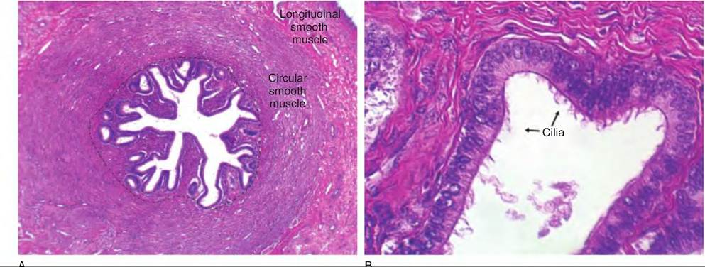
Fig. 19.1. Histology of the bovine female reproductive tract. Panel A shows a low power (4?) view of a cross section of the oviduct. The region outlined with the dashed line contains the mucosa and submucosa. Most evident in this view is the highly folded mucosal surface surrounding the opening of lumen of space in the center of the oviduct. The surrounding muscularis occurs as circular and longitudinal layers are evident as a band that surrounds the mucosa. A small edge of the serosa appears in the extreme upper right corner. The mucosa is enlarged (40?) in panel B. Here the epithelial cells appear as columnar epithelial cells with evident cilia.
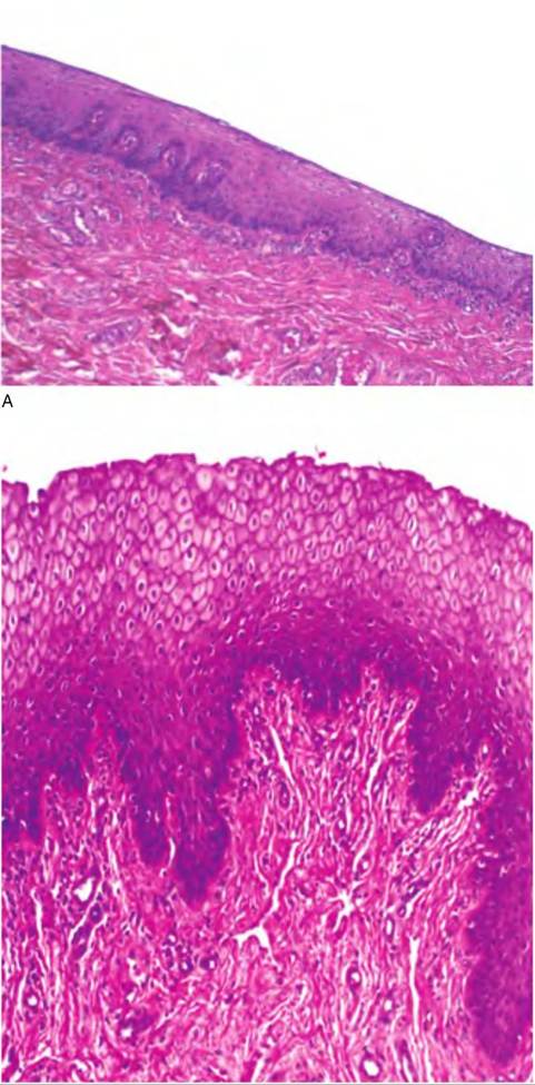
As you might suspect, there is substantial deviation in the surface epithelium at different locations of the reproductive tract. There are also marked variations associated with stage of the estrous cycle. For example, Figure 19.2 illustrates the vaginal epithelium along the surface of the posterior vagina as well as differences
B
Fig. 19.2. Posterior vaginal epithelium of bovine. During the luteal phase (A), the epithelial cell layer is thinner than during the follicular phase (B) of the estrous cycle.
associated with stage of the estrus cycle. Unlike the oviduct, here the epithelial lining is composed of stratified squamous epithelial cells. During the follicular phase of the estrus cycle, the layer of cells is thicker compared with the luteal phase.
For primates that express menses, variation in the uterine epithelium and in uterine glands is pronounced. Figure 19.3 shows the histological appearance of the uterus. Compared with other regions of the reproductive tract, the muscularis is extensive, and the submucosa area has abundant glands that supply secretions that are especially important during pregnancy.
The reproductive tract develops in a retroperitoneal position, so that it is positioned against the peritoneum. With continued growth, the tract becomes completely surrounded. This new connective tissue sheath forms a continuous drape around the reproductive tract. This functions to suspend and maintain the position of the ovaries, oviduct, uterus, and the anterior vagina. In this mature state this supporting tissue is called the broad ligament.
The ovaries, analogous to the testes in the male, are the primary reproductive organs since they produce the female gametes (ovum, singular, or ova, plural). However, the ovaries produce not only gametes; they are also critical endocrine organs. Release of mature
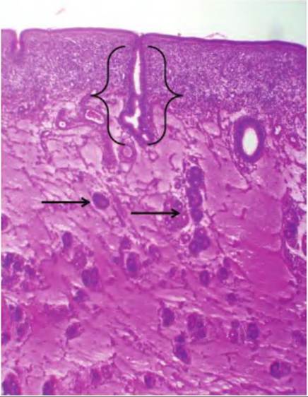
Fig. 19.3. Section of bovine uterus from a cow during the follicular phase of the estrus cycle (4?). A section through a uterine gland is evident (brackets) as well as older glands deeper within the muscularis (arrows).

Fig. 19.4. Gross anatomy of the female bovine reproductive tract.
ova occurs with the rupture of follicles on the surface of the ovary. The ovum or ova enter the funnel-like open end of the oviducts (infundibulum) to be directed to the uterus. Fertilization usually occurs within the oviduct during transit to the uterus. Figure 19.4 illustrates the gross anatomy of the primary structures of the reproductive tract of a cow and Figure 19.5 a dissected view of the vagina and cervix.
The paired ovaries are located in the lumbar region in close proximity to the kidneys. In sexually mature animals, the ovary undergoes dramatic but predictable cyclic development. For example, within a window of about 3 weeks in cows, pigs, or horses, ovulation occurs, and the selected ovarian follicles are transformed into corpus Iuteum (singular) or corpora Iutea (plural) that produce large quantities of progesterone.
If fertilization does not occur, the corpus Iuteum regresses, and a new crop of follicles matures. These follicles produce estrogen, and selected follicle(s) proceed to undergo ovulation. This pattern constitutes an estrus cycle. The ovary is oval to round in shape with distinct regional differences and blister-like structures—the follicles—near the outer surface. A connective tissue layer called the tunica albuginea covers and protects the ovary. This connective tissue band supports the surface layer of epithelial cell, which is unfortunately called the germinal epithelium. This is unfortunate because despite the promising name, these epithelial cells do not produce the gametes. Instead, beneath the tunica albuginea, within the ovarian cortex, there are populations of oocytes, which are recruited to develop into the mature follicles. The ovarian cortex also houses the corpus Iuteum as well as older degenerated corpora Iutea called corpora albicantia. The center of the ovary is called the ovarian medulla and contains the blood, nerves, and lymphatic vessels that supply the ovary. The structure of the bovine ovary is illustrated in Figure 19.6.In domestic species, the uterus consists of a body, a cervix (or neck), and two uterine horns. However, there are substantial variations in the shape and arrangement of the horns. For example, the body of the bovine uterus appears larger because the intercor- nual ligament, which acts to obscure the individual nature of the two uterine horns, covers the caudal region of the uterus. Among mammals there are three distinct types of uteri. The duplex uterus has two cervical canals, which act to separate each uterine horn into distinct compartments. However, there are two types of duplex uteri. In one there is a single vaginal canal opening to the outside. On the interior it divides to produce two vaginas and two cervices. This occurs in marsupials. In the North American opossum, for example, the male accommodates this circumstance by having a forked penis.
The rabbit has a less complex arrangement; there are two uterine horns and two cervical canals but a single vaginal canal. The bicornu- ate uterus is characterized by the presence of two uterine horns and a small uterine body. In all of these cases, the uterus opens into the vagina via a single cervical opening. Cows, mares, and pigs all have this type of uterine structure. Primates, on the other hand, have a simplex uterus. There is a large uterine body but essentially no uterine horns.The appearance of the internal lining of the uterus, the endometrium, varies during the estrous or menstrual cycles and during pregnancy. The tissue is also highly glandular with a rich blood supply. The epithelial surface is a simple columnar epithelium in the mare but stratified columnar epithelial cells in ruminants. In addition, simple branched tubular glands provide secretions—called uterine milk—that are especially important during estrus and pregnancy. In many animals the uterine glands are scattered throughout the endometrium. But in ruminants, the internal uterine surface is punctuated by caruncles that are not glandular. These mushroom-like caruncles provide sites for attachment of fetal membranes in these animals. The smooth muscle of the uterus (the muscularis) is frequently called the myometrium.
The most caudal region of the uterus leads to the cervix. The cervix is a tough, connective tissue and smooth muscle sphincter that is tightly closed except during estrus and parturition. During estrus, the slight
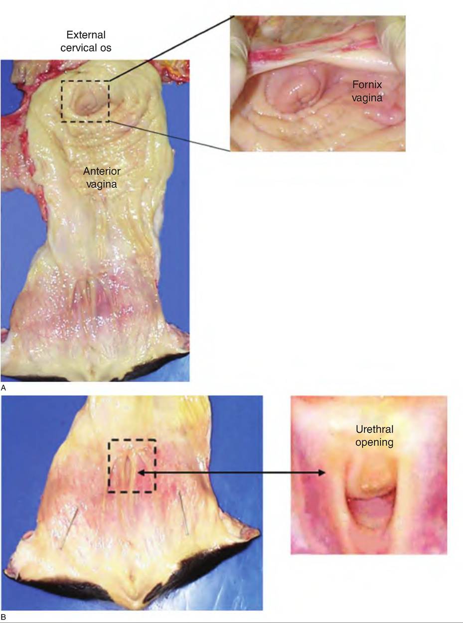
Fig. 19.5. A dissected view of the portions of the bovine vagina and cervix (panel A). The anterior vagina ends at the opening of the cervix (indicated by the dashed box, the external cervical os). The pocket on either side of the cervix (panel B) (fornix vagina) can be a nemesis for those learning the "art" of artificial insemination. Toward the rear or posterior end of the vagina (panel C), the opening to the urethra (panel D) is recessed in the suburethral diverticulum, an area that has to be successfully navigated for placement of urine cannulas used for experiments requiring collection and monitoring of urine production in cows.
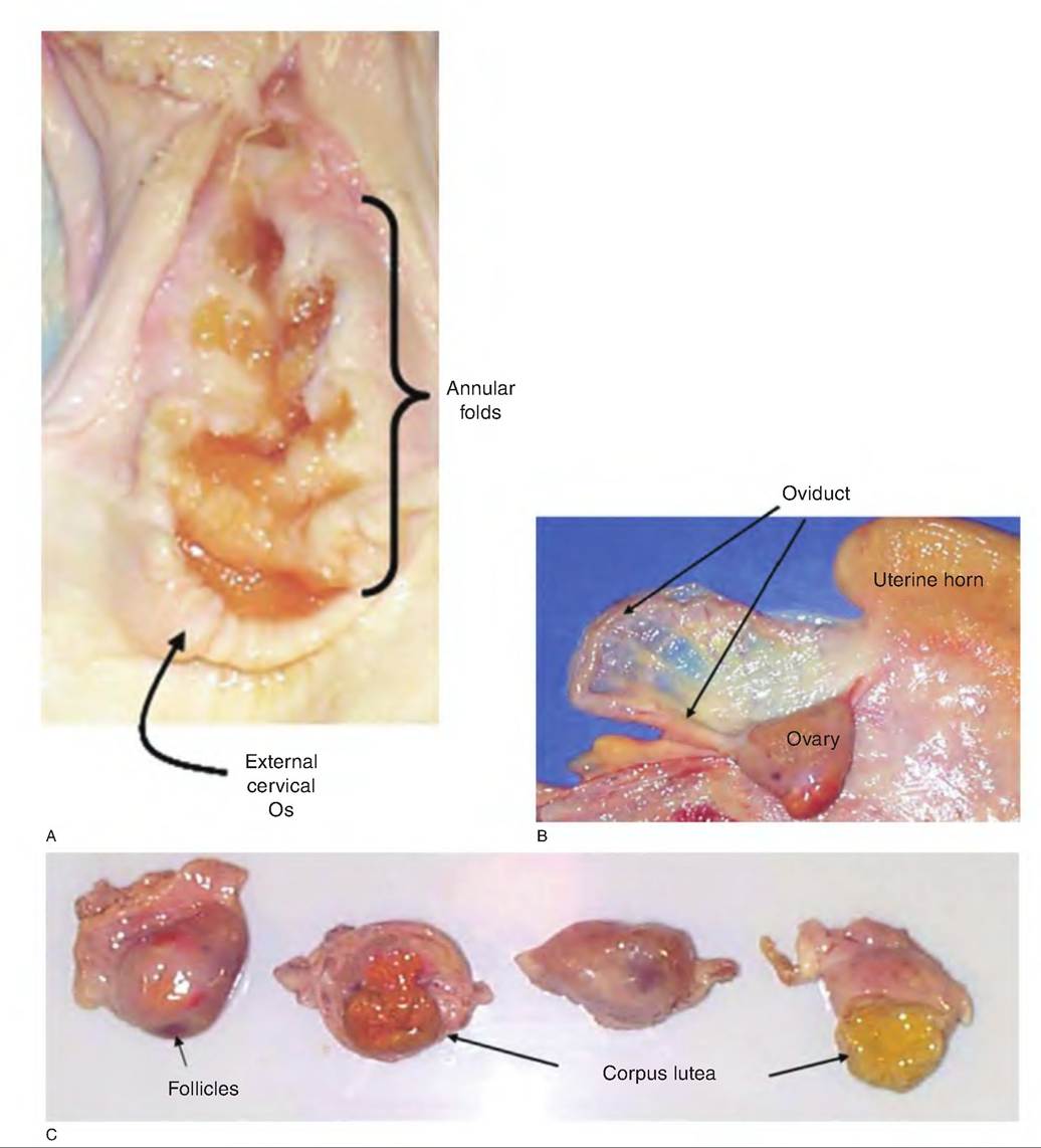
Fig. 19.6. Reproductive tract anatomy continued. Panel A shows a bisected view of the cervix. The circular pattern of the annular folds is evident. Panel B shows the relationship between the uterine horns, oviduct, and ovary. Panel C illustrates variation in follicles and corpus Iutea. The follicle to the left is nearing ovulation, as indicated by the red points in the surface. The third follicle is relatively mature (evidenced by the fluid-filled cavity [center reddish brown area]). The left corpus Iuteum is older than the more yellow body to the right of the panel.
| Table 19.1. Comparative anatomy of female reproductive tracts. | ||||
| Animal | ||||
| Organ | Mare | Sow | Cow | Ewe |
| Oviduct | 20-30 cm | 1 5-30 cm | 25cm | 15-1 9cm |
| Uterus | ||||
| Type | Bipartite | Bicornuate | Bipartite | Bipartite |
| Horn | 15-25 cm | 40-65 cm | 3 5-40 cm | 10-12 cm |
| Body | 15-20 cm | 5 cm | 2^fcm | 1-2 cm |
| Endometrium | Prominent longitudinal folds | Slight longitudinal folds | 40-120 caruncles | 88-96 caruncles |
| Cervix | ||||
| Lumen | Conspicuous folds | Corkscrew-Iike | 2-5 annular rings | Annular rings |
| Opening to uterus | Clearly defined | Ill-defined | Small and protruding | Small and protruding |
| Vagina | 20-35 cm | 10-15 cm | 25-30cm | 10-14cm |
| Vestibule | 10-12 cm | 6-8 cm | 10-12 cm | 2.5-3 cm |
Modified from Frandson et al. (2003).
loosening of the cervix allows spermatozoa to enter the uterus. In ruminants, the inner surface of the uterus is oriented in a series of circular folds or ridges called annular folds. Learning to traverse these folds can be a challenge to beginning artificial breeding technicians.
The vagina is the region of the reproductive tract within the pelvis positioned between the cervix on the cranial end and the vulva on the caudal end. The vulva or external genitalia is composed of the right and left labia, which join at the midline to produce a commissure or union. The ventral commissure of the vestibule (the posterior vagina) houses the clitoris, the female homolog of the glans penis in males. It contains erectile tissue and is covered by stratified squamous epithelium. It is well supplied with sensory nerve endings. The functional significance is not well established in domestic animals but clitoral stimulation at the time of insemination has been shown to increase conception rates in beef cows. Some comparative features of the female reproductive tract of various nonpregnant farm animals are given in Table 19.1.