PATHOGENESIS, PATHOLOGY AND IMMUNITY
Francisella tularensis is a highly infectious pathogen. It can enter the body in several ways: via inoculation by haemat- ophagous arthropods, through skin lesions, across ocular- mucous membranes, by inhalation of infected aerosols or by ingestion of contaminated meat (cannibalism) or water.
Only a small inoculation dose (LD50: 106 CFU) or humans (LD50: >103 CFU)(9).After entering the body the bacteria multiply locally, causing ulceration and necrosis, and then invade the blood and lymph vessels and spread to the lymph nodes and organs, including liver, spleen, lung, kidney, the serosal membranes and bone marrow, causing multiple foci of coagulative necrosis. Francisella tularensis is a typical intracellular pathogen with a high predilection for growth in macrophages but can infect many other cell types, such as epithelial cells, hepatocytes, muscle cells and neutrophils.
Francisella tularensis septicaemia occurs as an end stage of the disease, when the bacteria invade the blood vessels without lesions indicative of a tissue response. This septi- caemic form is seen in highly sensitive species and animals, which die within 2-10 days.
The pathology of tularaemia differs considerably between different animal species and gross lesions are not always observed. In Scandinavia, acute forms of tularaemia have been described in mountain hares(8), whereas in Central Europe infection of European brown hares apparently has a more chronic course(6). In mountain hares, the most characteristic necropsy finding is the enlarged spleen. Multiple white foci of coagulative necrosis can be seen in the spleen, liver and bone marrow in some, but not all, acute cases. Haemorrhagic enteritis and typhlitis can be found as well, particularly during winter. The mucosa in the jejunum and caecum is congested and occasionally necrotic.
The crypts and villi of the intestine may show focal necrosis. Histologically, the focal lesions are initially characterized by apoptosis, absence of inflammatory cell infiltrate and thrombosis of small vessels. In subacute cases the lesions are inflammatory and granulomatous, with necrosis and a proliferative reaction of macrophages and giant cells.In subacute cases in European brown hares, numerous, randomly distributed, well-demarcated, greyish-white or yellowish-white foci, with a diameter of 0.1 to 1 cm, can be observed most frequently in the lungs (Figure 22.1), the pericardia (Figure 22.1) and the kidneys(6). Foci are frequently observed on the serosal surfaces of the lungs and kidneys. These foci and those in the pericardium are raised, dome shaped or flattened, and their surfaces are dry and granular. Foci can sometimes be seen in the testicles, bone marrow and mammary glands. Gross lesions are usually not found in the spleen and liver. Histologically, the foci are randomly distributed and correspond to focal or coalescing granulomatous inflammation, which replaces the normal tissue structure. Macrophages are the dominant
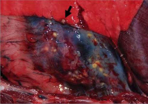
FIGURE 22.1 Pericardium and lung of a European brown hare. Note the numerous yellowish-white, flattened foci of different sizes on the pericardium and one greyish-white nodule surrounded by a dark, hyperaemic area in the lung lobe (arrow).
constituent cell type, but other cells — including lymphocytes, heterophil granulocytes, multinucleated giant cells and fibrocytes — are also found occasionally. Focal or multifocal necrosis is often observed in the centre of these lesions. Granulomatous inflammation can also be detected as microscopic lesions in the mediastinal lymph nodes, liver and spleen.
The pathology of tularaemia in rodents depends on the sensitivity of the species. The usual macroscopic finding is the enlarged spleen and, less frequently, the liver. Pinpoint white foci can be seen on these organs. Microscopically multifocal coagulation necrosis is characteristically found in the spleen, liver, lymph nodes, bone marrow and lungs. Karyolysis, pyknosis and the presence of inflammatory cells such as macrophages and heterophils are observed in less acute cases.
Little is known about the immune response of the host to F. tularensis. Cell-mediated immunity has long been believed to be critical for protection. The importance of humoral immunity is also now recognized. Synergy between antibodies, T-cell-derived cytokines and phagocytes appears to be critical to achieving immunity against F. tularensis and clearing infection. In humans, an antibody response is measurable by the second week post-infection. Antibody levels are highest during the second month after infection, and decline gradually thereafter.