PATHOGENESIS, PATHOLOGY AND IMMUNITY
Bacillus anthracis expresses its pathogenic action mainly through the capsule and the production of a toxic complex consisting of three proteins, protective antigen (PA), lethal factor (LF) and oedema factor (EF).
The toxic factors result from two plasmids: pXO1(182kb), carrying the genes encoding for EF, LF and PA and pXO2 (96 kb),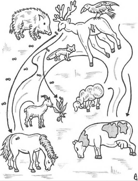
FIGURE 25.l Anthrax cycle in rural areas of Europe. This cycle involves wild animals and domestic animals. Wild animals die from anthrax and the carcass is opened by scavengers. This permits the sporulation of Bacillus anthracis. Spores are disseminated in the environment by carnivores, wild boar and scavenger birds. Blowflies infect directly receptive animals (horses and cattle). Contaminated water could contribute to the increase of the contamination level of pasture. Rainwater, having washed away the surrounding ground, tends to stagnate in low-lying parts, favouring the concentration of spores. Flies could contribute to environmental contamination. This sequence of events encourages the adhesion and distribution of spores on soil humus, increasing the chances of infecting herbivores.
carrying the genes encoding for the biosynthesis of the capsule. The PA binds to cell-surface receptors and plays a fundamental role in pathogenesis. It has no direct toxic action but acts as a ‘mover’ of the other two toxic proteins into cells. The capsule, a linear polymer of γ- D-glutamic acid, is considered the other major virulence factor of B. anthracis. The capsule contributes to pathogenicity by enabling the bacteria to resist phagocytosis by macrophages and thus evade the host immune defences and promote septicaemia.
The most frequent method of penetration by spores is via the digestive system after the ingestion of spore- contaminated feed, forages and water.
The portals of entry are micro-wounds that can be found in the mucous membranes of the mouth, pharynx and along the entire gastrointestinal tract. The infection can also occur through skin abrasions or skin lesions that may be caused by hae- matophagous insects (e.g. biting flies) acting as passive carriers or biological vectors. The severity of the disease depends on the sensitivity of the host, on the size of the infectious dose and on the route of penetration. It is considered that the spores of B. anthracis are carried by macrophages from the initial site of entry to the draining lymph nodes. The spores germinate, giving rise to vegetative forms that are capable of producing the main virulence factors: the toxins and capsule. These enter the bloodstream, where they continue to rapidly multiply. The pathogenicity of B. anthracis depends on the quality of the capsular coat and the amounts of toxins produced, and on the susceptibility of the host species.The principal lesions in septicaemic anthrax are widespread oedema, haemorrhage and necrosis. The nature and the extent of the lesions may vary and are dependent on the route of infection, host susceptibility and the virulence of the bacterium. Thus, in some carcasses, only lesions consistent with septicaemia are evident, whereas in others, localized necrotizing lesions are found.
I n ruminants, in which the course of the disease is usually acute or hyperacute, the most consistent changes include: evidence of rapid post mortem decomposition of the carcass, with marked bloating soon after death; incomplete development of rigor mortis; oozing of blood or blood-stained fluid from the natural openings such as the nose, mouth and anus; dark- r ed, poorly clotted blood; petechiae and ecchymoses throughout the body; extensive pulmonary oedema; excessive amounts of blood- tinged serous fluids in the peritoneal, pleural and pericardial cavities; and oedema and haemorrhage in individual lymph nodes. As suggested by the names ‘splenic disease’ and ‘miltsiekte’, severe splenomegaly is considered by many to be the most characteristic gross change, but it is not a consistent feature and its absence does not exclude anthrax. Most wild ruminants that die from anthrax manifest a vasogenic brain oedema coupled with the presence in the larger blood vessels of poorly formed and disintegrating post mortem blood clots, containing numerous encapsulated bacteria. Severe inflammatory oedema of the soft tissues of the head, tongue and throat, stomach and intestines are characteristic features of anthrax in carnivores. Bacillus anthracis septicaemia and bacteraemia are consistent features at death. Innate host resistance to infection by B. anthracis appears to be dependent on the inhibition of the initial multiplication stage of infection.