Sample-Specific Techniques and Challenges
19.5.1 Faeces
Faeces, as the primary test sample used for antemortem PCR-based diagnosis of paratuberculosis, is a non-invasive and easy sample to collect but is a difficult sample type to work with.
In addition, shedding can be intermittent and the levels of MAP present can often be low, depending on the stage of the disease. It is important in terms of disease diagnosis at the individual or herd level to be able to detect and monitor these low levels of MAP in the faeces. Early identification of individuals that are shedding the MAP bacterium prior to clinical disease onset is important for control, particularly as the majority of infected animals in any herd or flock are likely to be subclinical cases.Ruminant faeces is an extremely challenging sample due to the presence of the faecal microbiota (O' Donnell et al., 2017), as well as undigested particulate and plant matter, humic acid, bile and proteins, among other components. For ruminants, undigested residues of herbivorous matter such as cellulose, hemicelluloses and lignin constitute a major part of the faecal matrix. Many of these substances can be inhibitory in qPCR reactions, due to interactions with the components involved in amplification of the DNA, leading to potential false-negative results (Schrader et al., 2012).
It may not be achievable to predict and address the complex and variable nature of ruminant faecal samples given differences in breeds, production systems, diets, gut microbiome, health status, seasonal and regional variations. Thus, scientists in this area need to apply approaches to cope with this complexity in the local setting and validate these approaches. One quote that speaks to the trade-off between DNA purity and test sensitivity is ‘effective removal of faecal components (from DNA extracts) has to reach a balance between obtaining a concentration of inhibitors at a level that does not affect PCR amplification, and a concentration of target DNA sufficient for PCR amplification' (Rapp, 2010, p.
1489).Approaches to develop molecular detection assays for faeces that have high sensitivity and minimize the impact of inhibitors have been numerous. Debris present in the faecal sample is often allowed to settle out by gravity or gentle centrifugation and only the upper aqueous supernatant of the faecal suspension is included in the test (Sting et al., 2014; Mita et al., 2016). Immunocapture techniques have been applied as a means of reducing the carry-over of PCR inhibitors (Chui et al., 2010). Extraction kits that are designed to deal with complex samples, such as soil and faeces, have been utilized in the DNA extraction step for detection of MAP in faeces (Imirzalioglu et al., 2011; Arsenault et al., 2019).
A magnetic bead-based DNA purification method has been used during the extraction step by a number of researchers in order to achieve optimal PCR results and limit the negative effects of faecal components (Petrich et al., 2006; Plain et al., 2014; Sevilla et al., 2014; Schwalm et al., 2018). Rather than column-based methods, which require unbound sample lysate to be washed through the silica membrane, the advantage of this method is that the nucleic acids bind to the magnetic beads and are physically removed from the remaining sample lysate through successive wash steps in order to remove any contaminants. This type of extraction method has been applied in forensic testing of challenging environmental samples to improve the ability to obtain a genetic profile (Bogas et al., 2011).
Table 19.3 summarizes qPCR assays to detect MAP in faeces, including a broad range of DNA extraction approaches, that have been published in the past 10 years. The diagnostic sensitivity of these, compared with faecal
Table 19.3. Published studies on the development of direct faecal quantitative PCR (qPCR) assays to detect Mycobacterium avium subsp. paratuberculosis (MAP) in faecal samples, with information regarding analytical and diagnostic performance (Jan 2008 - July 2019).
| Author | Year | Species | No. samples (total) | qPCR method | Reference test(s) | ASea | ASp | DSe (No. samples) | DSp (No. samples) | ||||||||
| DNA extraction method | Bead beating (YZN) | Target gene(s) | Assay chemistry | ||||||||||||||
| Arsenault et al. | 2019 | Ovine | 77 | Spin columnb | Y | IS900' | Probe, two-step | TC, LJ FCd | Previous0 | Previous0 | 0.84 (n = 44) | 0.93 (n = 30)e | |||||
| (2019) | MAP DNA test kit (Tetracore) | Y | hspX | Probe, two-step | Com | Com | 0.52 (n = 44) | 0.93 (n = 30)e | |||||||||
| Schwalm et al. | 2018 | Bovine | 157 | Magnetic bead | Y | IS900= | Com | M7H9C | 0.2 GE | Com | 0.86 (n = 107) | 1.0(n = 50) | |||||
| (2018) | isolation1 | liquid FC1 | |||||||||||||||
| I Dveth | Com | IS900' | Probe, two-step | 0.2 GE | Com | 0.89 (n = 107) | 1.0(n = 50) | ||||||||||
| Mitaeta/. (2016) | 2016 | Bovinek | 24 | Five methods | Y | IS900l | SYBR Green, | HEYM FC | Previousm | Previousm | 0.71-1.0 (n = 24) | Previousm | |||||
| assessed1 | two-step | ||||||||||||||||
| Sevilla et al. | 2014 | Bovine | 615 | Magnetic bead | Y | IS900d | Triplsx (IAC), | HEYM and | 10-100 | 1.0 | 0.59 (n = 88), 0.70 | 0.85-0.94 (n = | |||||
| (2014) | isolation11 | ISMAP02 | probe, two-step | LJ FC | MAP/g | (n = 74)p | 453)e | ||||||||||
| Plain etal. (2014) | 2014 | Bovine | 1330 | Magnetic bead | Y | IS900 | SYBR Green, | BACTECtm | 10 MAP/g | 1.0 | 0.61 (n = 870)r | 0.996 (n = 460) | |||||
| Ovine | 596 | isolation4 | two-step | RM liquid FC | 0.84 (n = 507)r | 0.99 (n = 89) | |||||||||||
| Sting et al. | 2014 | Bovine | 116 | Eight methods | Y | IS900t | Probe, two-step | HEYM FC | 6 GE | nd | 0.59-0.85 (n = 116) | nd | |||||
| (2014) | assessed® | ISMav2u | Probe, two-step | 28 GE | nd | 0.46-0.65 (n = 116) | nd | ||||||||||
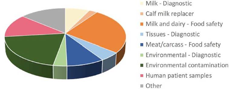
| Salgado et al. | 2013 | Bovine | 351 | PMSZethanoI | Y | IS900 | Probe, three-step | M G IT™ FC | TaqMan assay from Irenge et al. (2009);d infection status of culled sheep determined by TC, FC and histopathology;e population included animals from /WAP-exposed herds/flocks,f NukEx Mag Extreme extraction kit (Gerbion),9 diarellaMAP vet PCR (Gerbion),h IDvet Preparation kit/IDvet Extraction kit (IDvet Genetics);i IDGene™ Paratuberculosis duplex PCR (IDvet Genetics),i M7H9C media from Whittington etal. (2013);k pooled faeces not individual faecal samples;l methods: Johne-Spin kit (Fasmac, Japan), Tetracore MAP Extraction System (Tetracore lnc.), MagMAX™ Total Nucleic Acid Isolation kit (Applied Biosystems), QIAamp stool DNA Mini kit (Qiagen), ZR faecal DNA MiniPrep (Zymo Research); m previously published (Kawaji etal., 2007);n MagMAX™ total nucleic acid isolation kit (Ambion, Applied Biosystems);0IS900 assay from Herthnek and Bolske (2006);p based on follow-up testing of animals from two separate herds;9 modified method incorporating BioSprint 96 one-for-all vet kit (Qiagen);r more positives detected by qPCR than FC;s methods: protocol from Selim and Gaede (2012) or MagMAX™ Total Nucleic Isolation Kit (Life Technologies) ± four faecal suspension method variants;' IS900 assay based on Kim etal. (2002);u ISMav2 assay based on Wells etal. (2006);v study also includes 1293 human patient samples; w SmartHelix™ First DNAid kit (Institute of Physical Biology,Slovenia);x PSP Spin Stool DNA kit (Invitek);y proficiency kit samples;z Mississippi Veterinary Research and Diagnostic Laboratory (MVRDL) method, including chemical pre-treatment, cell lysis and extraction, chelex matrix absorption and spin column; α Johne-Spin kit (Fasmac, Japan);l3 QIAmp Stool Mini kit (Qiagen). MAP, Mycobacterium avium subsp. paratuberculosis; GE, genome equivalents; ASe, analytical sensitivity; ASp, analytical specificity; DSe, diagnostic sensitivity; DSP, diagnostic specificity; Com, commercial kit; FC, faecal culture; TC, tissue culture; LJ, Lowenstein-Jensen; HEYM, Herrold’s Egg Yolk Medium; RM, radiometric; IAC, Internal amplification control; PMS, peptide- mediated magnetic separation; ND, not determined. 312 K.M. Plainefa/. culture as the reference method, ranged from 0.46-1.0; this varied depending on the DNA extraction method, sample selection approach, target gene(s) and culture method used for comparison. Diagnostic specificity ranged from 0.85-1.0. However, true diagnostic specificity can be difficult to assess when the population studied includes MAP-exposed animals, as qPCR can be more sensitive than other diagnostic testing methods, often detecting additional positive samples compared with faecal culture. Further, PCR methods detect non-viable bacteria and some low positives may be due to passive shedding (Kralik et al., 2014b). Hence, the analytical specificity of the qPCR assay may provide a more accurate assessment of the likelihood of obtaining false-positive results. 19.5.2 Milk and dairy products Milk has been used as a test sample for both individual and herd-level diagnosis of paratuberculosis. A range of technologies can be applied: culture of milk or milk fractions, molecular methods (PCR, genotyping) and a rapid bacteriophage-based assay that specifically detects viable MAP, not just the presence of MAP DNA (Okura et al., 2012; Botsaris et al., 2016; Grant et al., 2017). The majority of recent publications of PCR to detect MAP in dairy products have been related to the food safety aspects for human consumption, as a potential zoonotic infectious transmission route, or calf milk replacers as a source of disease spread on-farm (Fig. 19.1, Table 19.4). The occurrence of MAP in milk on-farm has been previously reviewed (Okura et al., 2012). MAP is generally present in low concentrations in milk samples thus larger volumes are tested, with most methods incorporating a centrifugation step. Immunomagnetic or peptide- mediated separation prior to DNA extraction has also been used in order to ‘pull out' the MAP organisms from the sample prior to the test being conducted (Grant et al., 2002; Metzger-Boddien et al., 2006; Ricchi et al., 2014; Gilardoni et al., 2016). The analytical limit of detection of MAP in milk by PCR has been reported to range between 1 and 100 MAP/ml of milk, using qPCR methods with gene targets including IS 900, f57 ISMAP02 and ISMAV2 (Gao et al., 2007; Slana et al., 2009; Alajmi et al., 2016; Acharya et al., 2017b). The fraction of milk tested is significant; the majority of MAP is reported to be concentrated in the casein pellet rather than the whey (Acharya et al., 2017b). However, the cream layer can contain significant numbers of MAP and may also be important to consider in testing regimes (Gao et al., 2007; Herthnek et al., 2008). Molecular techniques applied to milk samples in the diagnosis of bovine tuberculosis may also be of relevance to the diagnosis of paratuberculosis (Lermo et al., 2010). PCR-based assays using milk as a sample may be best suited to the identification of highly
Fig. 19.1. Number of published studies by category using PCR methods to detect Mycobacterium avium subsp. paratuberculosis (MAP) in samples other than faeces (January 2008 - July 2019). Table 19.4. Examples of recent published studies on PCR assays to detect Mycobacterium avium subsp. paratuberculosis (MAP) by sample type, for samples other than faeces.
infectious animals. Stabel et al. (2014) reported the detection of MAP by PCR in 13% of milk samples from subclinical cows compared with 49% of samples from clinically infected cows, with viable MAP mainly found in milk/colos- trum collected during early lactation (≤60 days in milk). If used for herd-level surveillance, individual animal milk testing would require large numbers of animals to be tested to generate confidence in detection of low prevalence farms and is unlikely to be cost-effective. However, if the information obtained from the extracted DNA is utilized in another way, for example for epidemiological investigations (Kaur et al., 2010) or multiplex testing for a range of pathogens (Parker et al., 2017), then PCR-based methods may be suitable, otherwise they may be a more expensive test approach than a milk ELISA with similar diagnostic sensitivity. 19.5.2 Animal tissues and meat products Confirmation of infection with MAP at the individual animal level is commonly performed by culture of the MAP bacterium from the tissues of the intestine and mesenteric lymph nodes, which is considered the gold standard post-mortem diagnostic test for paratuberculosis. Molecular approaches can also be applied for detection of MAP in these same tissues, for rapid determination of the presence of MAP (Table 19.4). Challenges with detection of a pathogen in host tissues include the extraction method used and the high quantity of host DNA (Park et al., 2014). This is a common issue for molecular approaches attempting to identify a proportionally minute component of sequences specific for the pathogen in a high background of host DNA (Lee et al., 2017; Gu et al., 2019). High input quantities of DNA can cause inhibition of qPCR reactions and also interfere with the detection of the amplified product (Restrepo et al., 2006; Radomski et al., 2013). There are a number of ways to deal with this, such as target enrichment, simple dilution of the DNA extract and/ or optimizing the assay for input DNA quantity. Conventional PCR can provide an advantage over qPCR when applied to animal tissues as this method is less sensitive to the inhibition due to high levels of host DNA, thus researchers may choose conventional over qPCR for these applications (Zarei-Kordshouli et al., 2019). Validation is key to determine the limit of detection of the assay used. Tissue qPCR approaches may be of value in disease surveillance at slaughter or determination of farm/regional prevalence levels, as many current abattoir surveillance programmes involve examination of gross lesions and confirmation by histopathological and/or tissue culture, which is time-consuming and has relatively low sensitivity to detect infection (Munster et al., 2011; Acharya et al., 2017a). The majority of published studies on PCR-based detection of MAP in tissues relate to food safety or the detection of MAP DNA in human patient samples (Fig. 19.1). Methods applied in human research on Crohn's disease may utilize PCR due to anticipated low quantities of MAP in the intestinal tissue sample (Banche et al., 2015) (see also Chapter 3, this volume). Such PCR assays face the same issues as detection of MAP in animal tissues; greater cross-validation of PCR used in human and veterinary research would be of value to determine the analytical performance of these assays. 19.5.3 Other sample types Methods for detecting MAP by PCR have been published for samples as diverse as semen, blood, lymphatic fluids, environmental samples and culture medium (Juste et al., 2008; Munster et al., 2013; Khol et al., 2014; Plain et al., 2015; Hahn et al., 2017). MAP has been detected in the blood of animals with late-stage paratuberculosis by PCR and bacteriophage assays (Munster et al., 2013; Swift et al., 2013), although MAP detection in the blood is not a common finding at a herd or flock level (Bower et al., 2011; Bower, 2011; Swift et al., 2016). Detection of MAP in culture medium in order to confirm specific growth once posed a significant challenge, particularly for media containing egg (Whittington et al., 1998), though now commonly involves high-throughput approaches and/or commercial kits (Plain et al., 2015; Bauman et al., 2016; Rangel et al., 2017). Methods to detect MAP by PCR in slurry/ liquid manure, boot swabs, dust and other types of environmental samples have been published (Aly et al., 2009; Eisenberg et al., 2012; Hahn et al., 2017; Donat et al., 2019). These are informative to the mechanisms of disease spread on-farm as well as potential avenues for herdlevel diagnosis. The use of samples collected from the farm environment to identify infected herds is increasingly being investigated as an economical surveillance approach (Aly et al., 2009; Donat et al., 2016). There is also a substantial component of the literature regarding PCR to detect MAP in environmental samples that pertains to the risks and prevalence of MAP contamination of the environment (Fig. 19.1). A number of methods have been developed for water samples, for example to examine the effect of farm runoff on the environmental load in local waterways (Richardson et al., 2019). In this study, 1 l of water was filtered (0.2 μM) and the pelleted material extracted using a method incorporating bead-beating and a magnetic bead extraction kit, followed by qPCR detection of both the IS900 and F57 genetic elements. Water from rivers and tributaries in catchment areas within regions used for grazing by livestock (sheep and cattle) for both dairy farming and meat production exhibited up to 108 cell equivalents/l of MAP. 19.6
More medical literature on Medic.Studio
More on the topic Sample-Specific Techniques and Challenges:
-
Infectious diseases -
Internal diseases -
Obstetrics and Gynaecology -
Pediatrics -
Veterinary medicine -
-
Conflictology -
Ecology -
Economy -
Finance -
History -
Law -
Medicine -
Philosophy -
Religious studies -
| ||||||||