Sample stability
Fixed smears may be stored at room temperature for several days before staining and if ICC is requested, they can be fixed in methanol-acetone, wrapped in foil, and stored at −20°C.
Immunoreactivity is stable for several months. For flow cytometry, cells should be suspended in a liquid medium at a final concentration of 106 cells/ml, kept refrigerated, and processed within 24 hours. Some specific preservatives or freezing techniques may also be used, but immunoreactivity may be affected, therefore fresh samples are preferred. One or more cytologic slides should always accompany samples for flow cytometry, because they are necessary for correct evaluation of flow cytometric results. For molecular biology (e.g. PCR for analysis of T- and B-cell receptor gene rearrangements or detection of infectious diseases), tubes containing cell suspensions can be stored at −80°C. Also, stained or unstained cytology slides can be submitted for PCR analysis after being stored for extended periods of time.Cytologic features related to sampling
Often, FNAs of lymph nodes are unsuccessful and tissue surrounding or near the lymph node is aspirated instead. Mandibular nodes are in close proximity to salivary glands. Thus, aspiration of salivary epithelial cells is not uncommon (Figure 5.1). Samples from the salivary gland are cellular and viscous. They contain a proteinaceous (mucinous) background that causes windrowing of cells in the sample. Typically, most cells are glandular epithelial cells with abundant vacuolated cytoplasm and a small rounded nucleus with dense chromatin. Occasional clusters of ductal epithelial cells forming small tubular structures may be observed. These cells have scant, lightly basophilic cytoplasm, distinct cell junctions, and a round nucleus. Variable numbers of small lymphocytes may be seen if the lymph node was concurrently aspirated, but lymphoglandular bodies are generally scarce.
Neutrophils and other inflammatory cell types are not expected unless there is an ongoing disease process causing sialadenitis. Many lymph nodes (popliteal for instance) are surrounded by adipose tissue, so samples from these nodes frequently show a scarce cellularity and an abundant population of normal adipocytes (Figure 5.2). Sampling internal lymph nodes is usually ultrasound guided. Ultrasound gel may be found on the smear (Figure 5.3) and should not be confused with metachromatic/azurophilic granules or microorganisms. 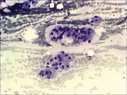
Figure 5.1 Dog, mandibular lymph node, FNA. Large clusters of glandular epithelial cells, likely from the salivary gland adjacent to the lymph node (May–Grünwald–Giemsa, 400? magnification).
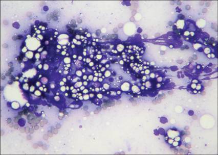
Figure 5.2 Dog, popliteal lymph node, FNA. Presence of lipid droplets and aggregate adipocytes from the adjacent adipose tissue intermingled with lymphoid cells (May–Grünwald–Giemsa, 600? magnification).
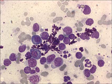
Figure 5.3 Cat, intestinal lymph node, FNA. Abundant purple granular material referable to ultrasound gel, among lymphoid and plasma cells from the reactive lymph node (May–Grünwald–Giemsa, 1,000? magnification).
Necrosis and disrupted cells are frequent if a very large lymph node is sampled.
Additional techniques to refine cytologic diagnosis