INTRODUCTION
During the last two decades, it has become evident that apoptosis plays an important role in a variety of physiological and pathological processes,1 including acquired immunodeficiency syndrome (AIDS).2 The growing awareness of the frequency and importance of apoptosis in these processes has resulted from an increased understanding of the biochemistry of apoptosis and concomitant development of methods for detecting this form of cell death.
Moreover, a critical understanding of the relevant literature requires knowledge of the strengths and deficiencies of the assays involved. In the sections that follow, we outline the biochemical basis for many of the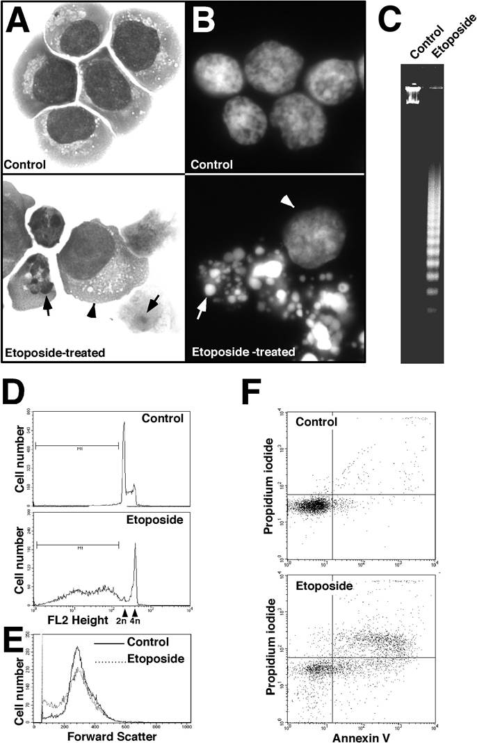
FIGURE 3.1 Examples of approaches used to detect apoptosis. (A) Detection by light microscopy. Jurkat human T cell leukemia cells were treated for 24 h with diluent (top) or 10 μM etoposide (bottom). Cytospins were prepared and stained with Wright’s stain. Compared to nonapoptotic cells (top panel and arrowhead, bottom panel), apoptotic cells are smaller and contain one or more nuclear fragments (arrows, bottom panel).
techniques used to detect apoptotic cells and then summarize the strengths and weaknesses of each approach.