Pathology of Insulin Signaling and Adipose Tissue
When adipose tissue is hypertrophic and resistant to insulin, as seen for visceral fat in metabolic syndrome, lipolysis is increased, resulting in increased levels of circulating free fatty acids (FFA).
An increased level of FFA plays a detrimental role on glucose utilization in the liver and muscles and leads to insulin resistance. The mechanisms responsible for this resistance to insulin can be hypothesized from the studies performed by Perseghin et al. [11] and are related to the inhibition of the insulin signaling pathway induced by acyl- CoA derivatives as indicated above. In addition, this excess of acyl-CoA inside the cytosol, exceeding the capacities of degradation in the mitochondria and peroxysomes, leads to accumulation of TG in hepatocytes and muscle cells. Such an accumulation has been reported in numerous studies of patients with insulin resistance, metabolic syndrome, obesity, and diabetes. Moreover, it has been consistently reported that the extent of fat accumulation in the liver is related to the amount of visceral fat and also to insulin resistance evaluated by HOMA or clamp tests [23]. Similarly, the amount of intramyocellular fat has been related to insulin resistance in obese and diabetic patients [24]. This set of alterations has been named lipotoxicity [25]. The location of adipose tissue is probably important; visceral fat, which is highly sensitive to catecholamines and resistant to insulin, is prone to release large amounts of FFA in the portal system which will be driven mainly to the liver. Subcutaneous fat, which is more resistant to lipolysis, would release lower levels of FFA, particularly towards peripheral tissues such as muscles.Metabolic syndrome, type 2 diabetes, and obesity are now considered as low- grade chronic inflammatory states, like atherosclerosis, leading to coronary heart disease.
Hyperinsulinemia and insulin resistance are considered as common preceding factors of hypertension, decreased HDL concentrations, hyper-TG, and altered glucose tolerance (Fig. 5). Part of the features of insulin resistance syndrome can be explained by the altered secretion of products from adipose tissue (and in particular from expanded visceral fat) with increased levels of proinflammatory cytokines, TNF-α and IL-6, responsible for this inflammatory profile. Interestingly, it has been shown in animal models of obesity and in human subcutaneous and visceral fat, that white adipose tissue presents macrophage accumulation, which is responsible for increased TNF-α expression [26—29]; increased endogenous production of cortisol increases insulin resistance participating in a vicious cycle (Fig. 6). Even if visceral fat is more difficult to study than subcutaneous fat in patients, it is highly probable that visceral fat is prone to release higher amounts of FFA and IL-6 and decreased amounts of adiponectin. They are released in the portal system and reach the liver first. This will result in increased production of acute inflammation proteins, such as CRP, and in increased glucose production and very-low- density lipoprotein (VLDL) synthesis resulting in hyper-TG [20].Subcutaneous abdominal fat, if insulinresistant, will produce increased FFA used by muscles, resulting in reduced glucose utilization at that level. The causes for this altered adipocyte function remain speculative: insulin can alter TNF-α and IL-6 secretion and, therefore, in the case of insulin resistance, these cytokines could be increased. Otherwise, insulin resistance could be a consequence of TNF-α and IL-6 oversecretion (Fig. 6). Increased levels of these cytokines at the level of the arterial wall lead to inflammatory lesions of atherosclerosis. Factors such as aging, smoking, and obesity can be responsible for increased proinflammatory cytokines [30]. This set of alterations will create a vicious
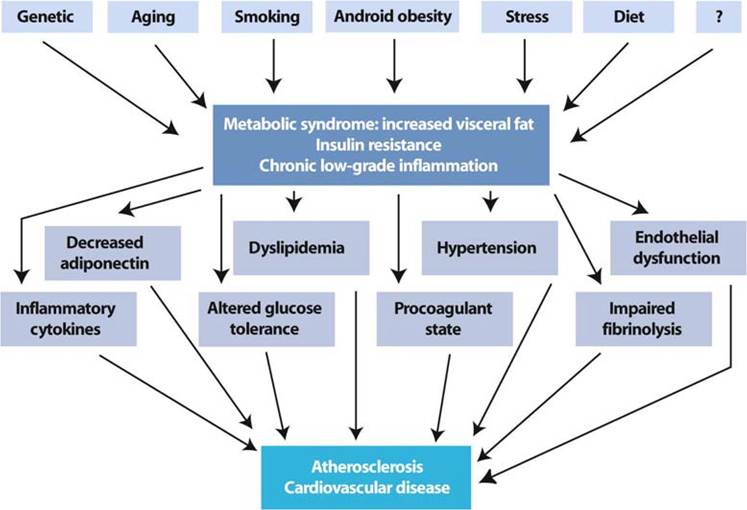
Fig. 5 Pathophysiology of metabolic syndrome
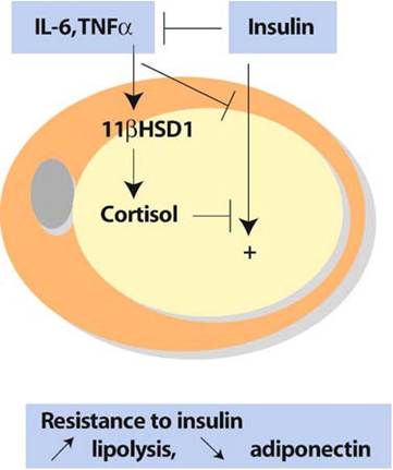
Fig. 6 Cytokines, cortisol, and insulin vicious cycle resulting in insulin resistance
cycle resulting in metabolic and vascular changes, ultimately leading to atherosclerosis and cardiovascular disease.