Endometrial carcinomas
Endometrial carcinomas present at a mean age of 63 years, however 4% of women with endometrial carcinoma are younger than 40, which has significant quality of life implications associated with childbearing and premature menopause related to treatment.
Almost all endometrial tumours present with abnormal uterine or postmenopausal bleeding and with prompt intervention they are frequently diagnosed in the early stage and have 5-year survival rates over 95% (2).Classification of endometrial carcinomas
In 1983, Bokhman classified carcinoma of the endometrium into two distinct groups (3). The first group described a group of tumours that originated in women with metabolic disturbances that include obesity, varying degrees of glucose intolerance, hyperoestrogenism and hyperlipidaemia. These tumours developed in a hyperplastic endometrium, were well to moderately differentiated, were less aggressive, and usually had a favourable prognosis. Tumours in Bokhmans second group were noted to be more aggressive, to have a greater chance of lymphatic spread, have higher recurrence rates, and the prognosis was less favourable than the first group. This dichotomous model has definite limitations due to heterogeneity within the two groups. Fortunately, advances in histological and immunohistochemical assessment of endometrial carcinomas have allowed these tumours to be more appropriately classified into high- risk and low-risk histological subtypes (Table 63.2).
The low-risk group incorporates tumours that have progressed through a stage of intraepithelial neoplasia (atypical endometrial hyperplasia) and so have maintained some degree of similarity to the parent tissue exhibiting varying degrees of endometrial gland formation and are thus described as endometrioid. The International Federation of Gynecology and Obstetrics (FIGO) provided a grading system of endometrioid carcinomas in 1988 that classified them into well (grade 1), moderately (grade 2), and poorly (grade 3) differentiated tumours, based on the progressive loss of glandular architecture leading to increasing solid growth patterns, as well as the degree of nuclear atypia.
Low-risk tumours include either well-differentiated or moderately differentiated endometrioid cancers and may exhibit genetic alterations including microsatellite instability and mutations in the PTEN, KRAS, and beta-catenin genes. They do not exhibit mutations in the TP53 gene. Poorly differentiated (grade 3) endometrioid carcinomas, while still being endometrioid by definition, bear little resemblance to endometrial glandular tissue. They have a greater chance of lymphatic spread, are more aggressive and often exhibit p53 mutation. For this reason, grade 3 endometrioid cancer is included in the high-risk group of endometrial cancers.The high-risk groups of tumours, while still originating from glandular epithelium, usually develop in an atrophic endometrium and display morphology that is structurally different from endometrial tissue. This group includes serous carcinomas and clear cell carcinomas. Both these exhibit TP53 gene mutations and chromosomal instability (4). Two additions to the high-r isk group include poorly differentiated (grade 3) endometrioid carcinoma and carcinosarcoma. Carcinosarcoma, which was previously classified
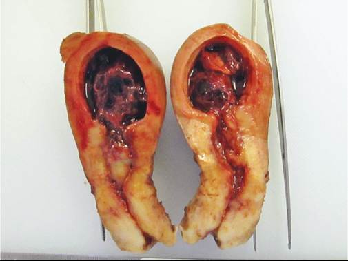
Figure 63.1 Uterine cancer.
as a uterine sarcoma, is now regarded as a carcinoma that has undergone metaplastic sarcomatous differentiation. It is a highly aggressive tumour with its carcinomatous component usually contributing to metastatic spread and is thus included in the high-risk group (5).
The majority of women with endometrial carcinoma have grade 1 and 2 endometrioid tumours and frequently present with early- stage disease and have excellent outcomes. The remaining 10-20% who are diagnosed with carcinomas of high-risk histology usually present in the late sixth and seventh decades. They often have more advanced disease and in over 50% of cases the disease has spread beyond the uterine corpus at the time of diagnosis.
Five-year survival rates in this group are significantly poorer and range between 40% and 60%.Risk factors
Risk factors for developing endometrial cancer can be divided into lifestyle factors, medical conditions, and hereditary factors. Medical conditions which are associated with the development of endometrial cancers are often sequelae of lifestyle factors, which are now becoming more evident in developing countries. Extreme body mass index, diabetes mellitus, hypertension, polycystic ovarian syndrome, early menarche, late menopause, infertility or nulliparity, unopposed oestrogen therapy, postmenopausal use of tamoxifen, oestrogen-secreting tumours, and HNPCC are all associated with
Table 63.1 Uterine malignancies
| Glandular Epithelium | Mucinous adenocarcinoma Serous adenocarcinoma Clear cell adenocarcinoma Carcinosarcoma |
| Supporting Endometrial Stroma/Mesenchymal cells | 1. Endometrial stromal sarcoma (previously lLow-grade endometrial stromal sarcoma) 2. Undifferentiated sarcoma (previously highgrade endometrial stromal sarcoma) 3. Adenosarcoma |
| Myometrium: | Leiomyosarcoma |
| Type 1 | Type 2 |
| Grade 1 endometrioid adenocarcinoma Grade 2 endometrioid adenocarcinoma | Grade 3 endometrioid adenocarcinoma Serous adenocarcinoma Clear cell adenocarcinoma Carcinosarcoma Undifferentiated carcinoma |
| Common genetic defects: KRAS, BRAF, PTEN, beta-catenin, mismatch repair defects (MMR) | TP53 |
an increased risk. The highest lifestyle-related risk is obesity (relative risk 2.54; 95% confidence interval 2.11-3.06) (6). Women who have inherited a HNPCC gene mutation have a 40-60% lifetime risk of developing endometrial carcinoma and a 9-12% risk of developing ovarian carcinoma due to defects in the DNA mismatch repair genes MLH1, MSH2, MSH6, and PMS2 (7).
A recent study has found that young patients with endometriosis have a 40% risk of developing endometrial cancer later on in life, the majority of these being type 1 (8). Pregnancy, use of the combined oral contraceptive pill, and cigarette smoking which either decrease oestrogen or increase progesterone levels have been shown to decrease the risk of endometrial cancer.Screening for endometrial cancer
Evidence does not support screening for endometrial cancer in a low-risk population as most cases cannot be avoided. There is no standard screening test. Ultrasonographic measurement of the endometrial thickness in low-risk asymptomatic women, may result in unnecessary interventions to sample the endometrium with potential procedure related complications and patient anxiety. To decrease the incidence of disease in the low-risk population, health professionals should rather focus on promoting healthy lifestyle choices, diagnosing and managing manifestations of the metabolic syndrome, and educating patients on the importance of having abnormal uterine or postmenopausal bleeding assessed.
Where tamoxifen is prescribed to postmenopausal women as part of breast cancer treatment, women should be counselled about the risk of endometrial polyps, hyperplasia, and endometrial cancer and that they should report any abnormal bleeding immediately. Tamoxifen has been reported to cause endometrial abnormalities in as many as 39% of women (9). The Mirena intrauterine system (IUS) has been used for endometrial protection. Although studies have not shown significant progesterone-associated risks of breast cancer development and carcinogenesis in breast cancer survivors with the use of Mirena, there are still concerns about oncogenic effects of any progestogens (10-12). Results from a small (n= 94) randomized controlled trial by Wong et al. could not demonstrate increased rates of recurrences nor breast cancer-related deaths amongst patients who were offered levonorgestrel as prophylaxis compared to non-users (13).
There is no good evidence to support the use of the levonorgestrel IUS (Mirena) as a preventative measure in these women (14).Women who are deemed high risk for developing endometrial cancer include carriers of the HNPCC mutation, women from families where there is a known mutation or where there is a strong
• Annual surveillance of the endometrium by gynaecological examination, transvaginal ultrasound, and aspiration biopsy starting at 35 years and should continue annually.
• Consider insertion of a levonorgestrel-releasing IUS.
• Prophylactic surgery using a minimally invasive approach should be discussed at the age of 40 years to prevent endometrial and ovarian cancer. The complications associated with surgical procedures as well as the uncertainty about the gains of prophylactic surgery should be discussed with honesty (15).
Endometrial hyperplasia
Endometrial hyperplasia is an abnormal proliferation of the endometrium and is driven by excessive levels of oestrogen, whether exogenous such as unopposed hormone therapy or endogenous from cyclical aromatization of androgens to oestrogens in excessive peripheral adipose tissue found in overweight women. A body mass index greater than 40 kg/m2 has been shown to increase the risk of atypical endometrial hyperplasia by 13- fold (9). Abnormal or uterine bleeding is the hallmark of presentation because of an unstable, proliferative endometrial lining. Endometrial sampling should be performed in women who present with abnormal bleeding over the age of 40 years, and in women younger than 40 with significant risk factors for endometrial cancer, including obesity and any suggestion or evidence of a hereditary mutation.
Histologically, endometrial hyperplasia is assessed architecturally as simple (ordered) or complex (disordered), and cytologically as being atypical or not. The most significant finding in the histological report is the presence of atypical cells, irrespective of the architecture as complex atypical hyperplasia is the immediate precursor lesion of endometrial carcinoma.
The risk of progression from complex atypical hyperplasia to invasive carcinoma is 29% compared to 8% in simple atypical hyperplasia and less than 3% in non-atypical hyperplasia (16). Atypical hyperplastic endometrium may already harbour a focus of invasive endometrioid carcinoma which may not have been sampled, especially when using blind sampling techniques. Differentiating between atypical hyperplasia and low-grade endometrioid carcinoma is often difficult and the opinion of an experienced gynaecological pathologist may be useful.Endometrial hyperplasia can be managed medically or with surgery. The choice of method is based on age, comorbid status, desire to retain fertility, and the presence of atypia. It is important to address potential sources of excessive oestrogen including obesity and to exclude oestrogen-secreting ovarian tumours in the management of endometrial hyperplasia.
Management of hyperplasia
Medical treatment
Endometrial hyperplasia without atypia can be managed medically using systemic or local progestins. Oral medroxyprogesterone acetate (10-20 mg daily) or megestrol acetate (160—320 mg daily) can be prescribed in either continuous or cyclical regimes. Response rates with medical therapy are good with positive responses seen after 3 months of treatment and complete response rates usually by 6 months to 1 year. Side effects of progestin therapy include weight gain, oedema, headaches, dizziness, and lack of libido are less common with local progestin therapy. The levonorgestrel-releasing IUS has been proven effective, and even superior, to oral medication in managing hyperplasia with and without atypia (17). Where there is no response to therapy after 6 months, progression to atypical hyperplasia, or unacceptable side effects, surgery should be considered.
Surgical management
In patients who are not medically fit to undergo surgery, progestin therapy must be prescribed (9). Endometrial hyperplasia where atypia is present is best managed by simple hysterectomy using minimally invasive techniques where possible. Bilateral salpingo- oophorectomy should be considered in postmenopausal women. Endometrial ablation and resection may be considered in women with complex non-atypical hyperplasia where medical therapy has failed and hysterectomy carries excessive risk or is against the patient's wishes. However, where atypical hyperplasia is present, ablative techniques are not recommended.
Clinical features of endometrial carcinoma
The most common symptom of endometrial carcinoma is abnormal uterine bleeding. The bleeding may be postmenopausal or in pre- and perimenopausal women it may be intermenstrual or prolonged bleeding. A thorough history, which includes a detailed menstrual history, recent cervical smear results, past and current use of hormone therapy, medical conditions, and family history of cancer and hereditary cancer syndromes must be taken. A full clinical examination should be performed which includes assessment of peripheral lymph node groups, examination of the breasts, abdominal examination, external and speculum examination of the lower genital tract, plus bimanual examination of the upper genital tract. Careful assessment of adnexal masses, uterine size and mobility, nodularity or masses in the pouch of Douglas, and parametrial disease are important to assess possible extrauterine spread and resectability.
Where no obvious cause of bleeding is detected on physical examination, the upper genital tract must be assessed using pelvic ultrasound scanning. Premenopausal women with significant risk factors for endometrial cancer and women over the age of 40 years must undergo histological endometrial assessment. There is no defined upper limit of endometrial thickness in premenopausal women, thus screening using sonographic endometrial thickness is not recommended. However, in premenopausal woman suspicion for hyperplasia or malignancy is increased when the endometrial thickness is greater than 12 mm. Office sampling using a flexible plastic endometrial sampler is adequate. A meta-analysis of sampling devices concluded that the Pipelle was the most sensitive device in detecting endometrial cancer (91—99%) and highly specific (98%) (18). In patients where office sampling is not successful, hysteroscopy is an excellent alternative and offers both visualization of the entire endometrial cavity and allows directed biopsy of any suspicious lesions.
In postmenopausal women, where bleeding is associated with an endometrial thickness greater than 5 mm, endometrial sampling must be performed. In women reporting a single episode of postmenopausal bleeding without significant risk factors, with a normal clinical examination and normal cervical cytology and an endometrial thickness less than 5 mm, sampling is not indicated (19). Studies have investigated the relationship between endometrial thickness and the risk of malignancy. Overall, using a cut-off value of 5 mm to perform endometrial sampling would offer satisfactory sensitivity with a false-negative rate of less than 1% (19). However, repeated episodes of postmenopausal bleeding, even where the endometrial thickness is less than 5 mm, must be histologically assessed as high-risk histological carcinomas develop in an atrophic endometrium (20). In such cases hysteroscopy may better assess an atrophic cavity. One study by Chandavarker et al. reported a diagnosis of either type 1 or type 2 endometrial cancer in 90 out of a total of 250 (36%) patients diagnosed with endometrial cancer, who had a prior endometrial stripe with a thickness not more than 4 mm (21).
Other less common presenting symptoms may include a pelvic mass, lower abdominal pain, and manifestations of metastatic disease including vaginal or vulval tumours or respiratory complaints. Not too infrequently, women may present to other disciplines with venous thromboembolic phenomena and on further investigation an endometrial malignancy may be diagnosed.
Cervical smears that report the presence of atypical glandular cells in an otherwise asymptomatic woman necessitate careful pelvic, colposcopic, and endometrial assessment. The presence of endometrial cells on a cervical smear in asymptomatic postmenopausal women also warrants investigation.
Further investigations following a diagnosis of endometrial cancer
Histological specimens reporting endometrial cancers or atypical hyperplastic endometrium, where possible, should be evaluated by an experienced gynaepathologist. Subtle differences in the continuum between atypical hyperplasia and endometrial cancer as well as the assessment of the grade of differentiation may not always be clear. Appropriate immunohistochemical staining techniques may also be required to differentiate between primary endocervical adenocarcinomas and endometrial adenocarcinomas and between uterine and ovarian serous carcinomas.
Biochemical and radiological preoperative investigations include the following:
• Full blood count.
• Liver function tests, specifically alkaline phosphatase and gammaglutamyl transferase.
• Urea, electrolytes, and renal function tests.
• Random blood glucose (to exclude undiagnosed diabetes mellitus).
• CA125 (has been shown to correlate with extrauterine spread in high-grade tumour but a consensus opinion is that there is no evidence to support the use of serum tumour markers in endometrial cancer) (14, 22).
• Imaging (dependent on local resources, institutional indications for staging lymphadenectomy, and whether fertility preservation is a treatment goal):
■ Pelvic ultrasound to determine tumour size, assess adnexa, cervical involvement, and depth of tumour invasion into the myometrium.
■ Pelvic magnetic resonance imaging (MRI) to size tumour and assess pelvic spread, myometrial invasion, cervical involvement, and pelvic lymphadenopathy
■ Computed tomography (CT) scan of chest, abdomen, and pelvis to investigate distant metastases to lung and liver, exclude hydronephrosis, assess peritoneal spread, and pelvic, para-aortic, mediastinal, and hilar lymphadenopathy.
■ In low-income countries, a chest X-ray and abdominal ultrasound are often the only available and affordable options.
■ CT-positron emission tomography, where available, has a high specificity but low sensitivity in excluding lymph node disease.
2009 FIGO staging of endometrial carcinoma
Endometrial cancer is staged surgically. In 2009, the Gynecological Oncology Group of FIGO published a revision of the 1988 staging system of endometrial carcinoma. This new system is felt to be a more accurate staging instrument and thus a better prognostic tool than its predecessor (Table 63.3) (23).
Surgical staging includes removal of the uterus and adnexae. A systematic pelvic with or without para-aortic lymphadenectomy should be performed in patients with high-risk tumour features such as grade 3 differentiation, non- endometrioid histology, cervical stromal invasion, lymphovascular system space invasion (LVSI), and where the tumour invades more than 50% of the myometrial thickness.
Cytological examination of peritoneal washings or free abdominal fluid is reported separately and does not upstage the woman. Malignant cells in peritoneal fluid have not been shown to be of prognostic significance (24).
Treatment of endometrioid endometrial cancer
The primary management of endometrial cancer is surgery.
Surgery
Surgery in endometrial cancer aims to remove the primary tumour, to perform comprehensive staging, and, in advanced abdominal disease, to achieve optimal cytoreduction where feasible. Surgery with curative intent may not always be possible where:
• there are patient-related factors such as extreme cachexia, extreme obesity, severe cardiorespiratory compromise, or recent acute pulmonary embolism or venous thromboembolism
• there is unequivocal (cytological or histological) evidence of parenchymal liver or lung metastases as well as lymph node involvement
• clinical or radiological examination suggests that pelvic or abdominal disease is not resectable.
Prior to implementing any form of treatment, each patient must be individualized:
• Fertility desires and surgical menopause in women under the age of 40 years must be discussed (see ‘Fertility-sparing options').
• Suitability for surgery must be assessed. Elderly, frail women and women with severe comorbidities may require assessment and optimization by a physician, geriatrician, and anaesthetist.
• The need for systematic lymphadenectomy must be determined. This may be determined by preoperative factors or by intraoperative assessment of the tumour (see ‘Lymphadenectomy in endometrial cancer').
| Stage I | Tumour confined to the corpus uteri |
| IA | No or less than half myometrial invasion |
| IB | Invasion equal to or more than half of the myometrium |
| Stage II | Tumour invades cervical stroma, but does not extend beyond uterus |
| Stage III | Local and/or regional spread of the tumour |
| IIIA | Tumour invades the serosa of the corpus uteri and/or adnexae |
| IIIB | Vaginal and/or parametrial involvement |
| IIIC | Metastases to pelvic and/or para-aortic lymph nodes |
| IIIC1 | Positive pelvic nodes |
| IIIC2 | Positive para-aortic nodes irrespective of pelvic node status |
| Stage IV | Tumour invades bladder and/or bowel mucosa, and/or distant metastasis |
| IVA | Tumour invasion of bladder or rectal mucosa |
| IVB | Distant metastases, including intra-abdominal metastases and/or inguinal lymph nodes |
Source data from 2009 FIGO staging of endometrial carcinoma.
• The extent ofsurgery must be tailored to each woman. Laparoscopic and vaginal routes may need to be considered. A shorter surgical time and less morbid surgical route may be necessitated by co- morbid disease or cardiorespiratory compromise. Intraoperative limitations in access or visibility due to visceral and abdominal wall adipose tissue may also make the procedure unnecessarily challenging. In certain cases, systematic lymphadenectomy, though indicated, may need to be abandoned.
Conventional surgery should include:
• a midline abdominal incision for non-endometrioid subtypes or where preoperative investigations suggest upper abdominal disease
• careful and bloodless entry to the peritoneal cavity
• peritoneal washings or sampling of free fluid
• abdominal survey exploring the entire peritoneal cavity in a systematic manner looking for tumour deposits and enlarged lymph nodes
• extrafascial hysterectomy with or without a vaginal cuff
• bilateral salpingo-oophorectomy
• removal of all suspicious or enlarged pelvic and para-aortic nodes
• infracolic omentectomy, to be considered in cases with serous histology (14)
• systematic lymphadenectomy, where indicated by institutional policy, based either on preoperative or intraoperative tumour assessment
• a maximal effort at cytoreduction (where feasible) has been shown to improve progression-free and overall survival rates (25).
Further surgical considerations include the following:
• Obesity is a major risk factor for endometrial cancer and a lower transverse incision is more suited to patients with clinically early- stage, low-grade disease. This group of patients seldom require systematic lymphadenectomy and rarely have extrapelvic disease. Wound complications, incisional hernias, and complications related to impaired mobility are less.
• Laparoscopic surgery: laparoscopy offers great benefit to obese women. It has been extensively researched and is widely used to manage endometrial cancer. For laparoscopy to be an effective alternative, it needs to be as safe and have equivalent oncological outcomes to open surgery. The Gynecologic Oncology Group (GOG) LAP2 trial showed that intraoperative surgical complication rates were similar between the patients who had laparotomy and those who had the laparoscopic approach. The laparoscopic group had significantly less minor postoperative complications and most women were discharged 2 days after surgery (26). A follow-up study of the LAP2 trial and a 2009 meta-analysis of trials comparing open and laparoscopic surgery found that the laparoscopic route did not significantly disadvantage oncological outcomes (27, 28). Retrospective studies have shown robotic surgery to have equivalent oncological outcomes to a laparoscopic approach (29).
• Routine radical hysterectomy for overt stage II tumours is not of survival benefit and increases intraoperative and postoperative complications (14). Women with stage II disease will receive adjuvant local radiation and any residual microscopic parametrial disease will be sterilized. Removal of the parametrium should only be considered when there is palpable parametrial spread and radical surgery will achieve clear margins.
• Synchronous ovarian tumours are a rare occurrence and more common in younger women. They are frequently of endometrioid histology and to decide whether they are metastatic spread from a primary endometrial tumour or a de novo ovarian carcinoma is challenging. In a study of17 women under 45 years with endometrial cancer, 29.4% had synchronous ovarian tumours compared to 4.6% in women over 45 years (30). In these cases, ovarian preservation is not appropriate. Each tumour site needs to be viewed as an independent cancer and managed with the appropriate surgical and adjuvant therapies.
• A vaginal approach should be offered in women fit for anaesthesia, but where abdominal surgery is not safe or achievable. Hysterectomy and bilateral adnexectomy should be possible using this route.
• Oophorectomy in young women: there is significant morbidity and mortality associated with removal of ovaries in young women. Not performing bilateral oophorectomy in those under 45 years without a significant hereditary risk and with grade 1 early-stage endometrial disease has not been shown to impact overall survival. Salpingectomy should still be performed (14).
• Palliative hysterectomy should be considered, where safe and feasible, in advanced disease to alleviate vaginal bleeding, offensive discharge, and pain. Patients with carcinosarcoma frequently present with distant metastases but also have a rapidly enlarging uterus often with a fungating and necrotic tumour mass protruding through the cervix. In selected patients, quality of life can certainly be improved by removing the uterus.
Lymphadenectomy
The main purpose of lymphadenectomy in endometrial cancer is surgical staging and should be considered in women with intermediate (stage I low-grade tumours with >50% invasion or grade 3 tumours with <50% invasion) or high-risk factors. However, lymphadenectomy also provides important prognostic information and is essential in evaluating the need for adjuvant therapy.
As with all gynaecological cancers, suspicious or enlarged lymph nodes detected at surgery should be removed, or at least sampled if removal is deemed unsafe (i.e. a node densely adherent to a major vessel). Systematic lymphadenectomy is probably the most controversial topic in the management of endometrial cancer. There has been much debate in terms of the indications, extent, and therapeutic and survival benefits of systematic lymphadenectomy.
As the majority of women with endometrial cancer present with early-stage low-grade disease, hysterectomy and bilateral salpingo- oophorectomy is sufficient treatment to achieve cure. Two randomized trials have shown that performing a routine systematic lymphadenectomy on these women offers no survival (31, 32). Such practice is unnecessary and increases intra- and postoperative complication rates. A 2015 European Consensus Conference strongly recommends against performing systematic lymphadenectomy in these women (14). However, the studies on which the current rationale for lymphadenectomy is based have been criticized for a number of reasons including:
• low node yields
• not including or only sampling the para-aortic nodes
• most study participants had early-stage low-risk cancers where lymphadenectomy would not have been of benefit
• a heterogeneous population in terms of tumour characteristics combining endometrioid and non-endometrioid subtypes and grade 1 and 2 with grade 3 tumours
• women were randomized to adjuvant radiotherapy regardless of the results of the lymphadenectomy.
Indications for a lymph node dissection
Systematic lymphadenectomy is performed based on specific indications. These indications try to exclude early-stage low-risk tumours where lymphadenectomy would be of no benefit. The indications have been determined using the results of various trials on endometrial cancer and they may be based on both preoperative and intraoperative factors. Exact indications for systematic lymphadenectomy are often institution dependent and are determined by resources and personal interpretation of controversial and contradicting data (Table 63.4).
Certain intraoperative factors are established using frozen section. This requires the presence of a gynaepathologist in theatre which
Table 63.4 Indicators for systematic lymphadenectomy
| Intraoperative | |
| Assessed using endometrial histology and imaging techniques such as pelvic ultrasound, MRI, or CT | Determined by intraoperative sectioning or frozen section (preferably by an experienced gynaepathologist) |
| • Non-endometrioid histology • Grade 3 differentiation • Visible cervical involvement • Radiological suggestion of deep myometrial invasion (>50%), cervical stromal invasion, lymphadenopathy >10 mm • Radiological measurement of greatest tumour surface dimension >2 cm (24) | • Upgrade of original grade to grade 3 differentiation • Extrauterine tumour • Non-endometrioid histology • >50% myometrial invasion • Cervical stromal involvement (Assessing grade of differentiation and myometrial invasion may be difficult on frozen section, especially in the presence of adenomyosis) |
may not always be possible, especially in developing countries. As an alternative, intraoperative sectioning of the uterus (preferably in the coronal plane) with visual assessment of the depth of myometrial invasion has been shown to be up to 87% accurate in grade 1 tumours and 65% accurate in grade 2 tumours (33, 34). Where the intraoperative tumour diameter is less than 2 cm, nodal metastases are unlikely (35). This practice may be of benefit where assessing the need for systematic lymphadenectomy via preoperative imaging or frozen section is not always available.
Extent of lymphadenectomy
In women where lymphadenectomy is indicated, there is no doubt of the need for complete resection of all pelvic nodes up to and including the common iliac nodes; however, the need for and extent of para-aortic dissection is controversial.
Complete para-aortic lymphadenectomy significantly increases surgical time, blood loss, surgical incision size, and postoperative complications including a 20% risk of lower limb lymphoedema (36). Women may be obese, frail, and often have significant comorbid conditions and the risk-benefit profile of such extensive surgery needs to be carefully assessed. The single factor that seems most predictive of para-aortic nodal involvement is confirmed pelvic node metastases. Both a Japanese study and one conducted by the Mayo Clinic (United States) have shown nearly 50% of patients with positive pelvic nodes have positive para-aortic nodes, whereas over 95% of patients had negative para-aortic nodes when the pelvic nodes were negative (37, 38). There may be uterine lymph channels that drain directly to the para- aortic nodes at the level of the renal vessels. This is explained by the collateral uterine-ovarian lymphovascular supply and accounts for the small group of women (1.6%) who have para-aortic metastases with negative pelvic nodes, where these positive nodes would be missed by pelvic lymphadenectomy alone (39).
Sentinel node biopsy in endometrial cancer
Following the success of sentinel node biopsy in breast and vulval cancers, sentinel node studies have been performed in endometrial cancer. This practice has the potential to afford every case of endometrial cancer a nodal assessment independent of risk factors. Performing sentinel node biopsy has been shown to detect lymph node metastases in 10% of women who would not usually have undergone systematic lymphadenectomy based on tumour factors (40). Its benefits include less surgical morbidity, specifically lymphoedema, while still providing essential information to stage the disease and tailor adjuvant therapy. There is some controversy about where the dye or fluorescent stain should be injected in endometrial cancer: be it at the cervix, as done when mapping sentinel nodes in early-stage cervical cancer, or via hysteroscopy into the uterine fundus or into the tumour.
Radiotherapy in the management of endometrial cancer
Radiation can be used for a number of indications in endometrial cancer including:
• adjuvant therapy for intermediate or high-risk disease
• primary treatment where surgery is not suitable
• treatment of recurrent disease
• palliation of symptoms including bleeding or symptomatic metastases.
While adjuvant radiotherapy has no significant effect on overall survival it has been shown to increase progression-free survival by reducing pelvic recurrences (32, 41, 42). The need for adjuvant radiotherapy in endometrial cancer is based on specific risk factors in stage I disease but is always indicated in stage II disease and greater.
Adjuvant radiation can be delivered in four forms: vault brachytherapy, external beam pelvic radiation, extended field radiation (to expand the irradiated field to include para-aortic nodes below the renal arteries), and whole abdominal radiation. The vagina is the most common site of recurrence. Vault tumours are difficult to manage surgically but vault brachytherapy and external pelvic radiation are effective and well tolerated adjuvant interventions (41, 43). Whole pelvis irradiation has been shown to be as effective as vaginal vault brachytherapy in reducing tumour recurrence at the vault; however, vaginal brachytherapy is preferred as the total radiation dose is smaller thus reducing radiation-mediated toxicity (42).
Surgery followed by adjuvant pelvic radiation was shown in the ASTEC study to reduce recurrence in the pelvis and at the vault in early-s tage disease but demonstrated no improvement in overall survival (32). The findings of the ASTEC study were mirrored in the PORTEC-1 study; however, both studies received much criticism (41). ASTEC had major design faults and the PORTEC-1 study included participants who were considered at low risk of developing recurrent disease.
PORTEC-2 randomized patients with an intermediate-risk profile for local recurrence to either vault brachytherapy or whole pelvis irradiation. No significant difference in vaginal vault recurrence or overall survival was seen in either arm; however, external beam treatment increased long-term morbidity (42). The findings of both PORTEC studies were criticized as lymphadenectomy was not performed in either trial, both were underpowered, and included large numbers of low-risk tumours that had minimal risk of pelvic nodal involvement and recurrence.
In early-stage low-risk tumours (stage I, grade 1-2, <50% myometrial invasion, LVSI negative) vault recurrence is not common and adjuvant radiotherapy is not indicated (14). In stage I disease with high- to intermediate-risk factors (grade 3 endometrioid with <50% myometrial invasion, or grade 1-2 with extensive LVSI) or high-risk factors (grade 3 endometrioid with ≥50% myometrial invasion regardless of LVSI) there is unanimous support in the literature that such cases require adjuvant radiation to decrease local recurrence; this was further supported by the GOG-99 study (44). In stage II disease where the tumour has invaded the cervical stromal tissue, a vaginal brachytherapy boost is often prescribed in addition to external beam radiation. In cases of high-intermediate-risk disease where the nodal status is unknown, adjuvant external beam therapy should be chosen above vault brachytherapy where there is unequivocal LVSI (14).
Primary treatment of endometrial cancer using radiotherapy is reserved for patients not suited to surgery. Radiation is delivered to the uterus using intracavity brachytherapy via a rod or Hayman's pack that is inserted transvaginally into the cavity. Intracavity therapy is usually combined with external beam radiotherapy, however, external radiotherapy may prove problematic as the weight and abdominal girth of certain patients may exceed the limits of the external beam radiation machine.
Chemotherapy in endometrial cancer
Chemotherapy in early-stage endometrial cancer
The role of chemotherapy in early-stage endometrioid endometrial cancer has not been established, however, chemotherapy may be beneficial in patients with early- stage carcinosarcoma. The toxicity of combination chemotherapy and only a marginal increase in overall survival may not justify use of these drugs (45, 46). Ifosfamide-based combination treatment, especially when combined with paclitaxel, has shown more activity in the treatment of advanced stage or recurrent carcinosarcoma (47).
Chemotherapy in advanced-stage disease
The combination of postoperative radiation and chemotherapy seems to give better progression-free and overall survival outcomes compared to either chemotherapy or radiation given as stand-alone treatments (48). Although multiagent chemotherapy is more effective, the cumulative toxicity is not well tolerated (49). In a GOG study, by Randall et al., chemotherapy seemed to offer better progression-free survival in stage III patients, with gross residual disease and endometrioid histology compared to whole abdominal irradiation (50).
Role of targeted therapies
Phosphotidylinositol-3 kinase (PI3K) is mutated in both type 1 and 2 uterine cancers. Current treatments aim at inhibiting this pathway (51).
Targeted therapies such as PI3K/AKTZmammalianZmechanistic target of rapamycin (mTOR) and vascular endothelial growth factor inhibitors are considered in oncological practice to:
• act synergistically with current chemotherapeutic agents
• reduce resistance to chemotherapy
• increase progesterone receptor expression in patients with advanced-stage or recurrent uterine carcinoma resistant to progestogens (52).
Most of these target-based treatments are being tested in phase II studies.
Metformin in endometrial cancer
Metformin is a biguanide that not only counteracts insulin resistance, but has been found to counter proliferative, invasive, and metastatic activity in patients with type 1 endometrial cancer. The antitumour effect of metformin is believed to inhibit the mTOR protein kinase (53).
Fertility-sparing management in endometrial cancer
Increasingly, more women under the age of 40 years are diagnosed with endometrial cancer. Fertility is sometimes a concern at the time of diagnosis. Most of these women are diagnosed with low-risk carcinomas or endometrial hyperplasia, though high-grade (grade 3) endometrioid carcinoma may be found in about 8% of young women (54). A high-stage endometrial cancer diagnosis is also not uncommon. The depth of tumour invasion should be considered before conservative treatment is considered. Dynamic MRI with contrast is more sensitive in assessing depth of tumour infiltration (55). Hormonal therapy has been used with reasonable resolution of disease. There have also been reports of successful pregnancies after hormonal treatment for endometrial cancer (56). Progression of disease has been observed in 47% of these women. Physicians should have a thorough discussion of possible risks and benefits of the conservative management before embarking on this approach.