Fetal risks
Fetal loss/miscarriage
There is a higher risk of fetal loss and miscarriage (pregnancy loss before 24 weeks' gestation) in multiple pregnancy, with risks increasing with the number of fetuses.
The higher fetal loss rates are mainly explained by the increased risk of poor implantation, fetal abnormality (aneuploidy and structural), extreme preterm labour, and in monochorionic twins, complications of shared placenta. One study estimates about 12% of all early conceptions start as twins (36). Resorption of one pregnancy in a previously ultrasonographically confirmed twin pregnancy, ‘the vanishing twin' phenomenon, occurs in up to 20% of first-trimester twin pregnancies. This has implications in first-trimester screening (24).Fetal abnormality
The risk of fetal anomaly is greater in twin compared with singleton pregnancy (37). The rate per fetus in dizygotic twins is probably the same as that in singletons, whereas it is two to three times higher in monozygotic twins, being attributed to abnormal cleavage in the latter. In around 1 in 25 dichorionic, 1 in 15 MCDA, and 1 in 6 mono amniotic twin pregnancies, there is a maj or congenital anomaly that typically affects only one twin (38, 39). Common structural abnormalities in twins include neural tube defects, anterior abdominal wall defects, facial clefts, brain abnormalities, cardiac defects and gastrointestinal anomalies. All multiple pregnancies should have a detailed scan for screening for structural defects, considering that scanning in multiples may take longer than singletons (40). NICE recommends that the ultrasound scans in twin and triplet pregnancies at a slightly later gestational age than in singleton pregnancies. Any discordant anomaly in twins warrants a referral to a tertiary fetal medicine unit (11). Even in monozygotic twins, concordance for a structural anomaly is found in fewer than 20% of cases.
One to two per cent of twin pregnancies will have a discordant anomaly, needing specialist assessment and counselling about the options of expectant management and selective reduction.Selective reduction in twin pregnancy
Selective reduction is an option for multiple pregnancy with discordant anomaly. This is usually offered for a major congenital abnormality or an aneuploidy. The procedure and the timing depend on the chorionicity, the gestation at which a discordant abnormality is picked up as this influences the risk of miscarriage, and/or preterm birth. Such cases should be referred to a tertiary fetal medicine unit with experience in such procedures. In dichorionic twins, a selective feticide is performed by ultrasound-guided intracardiac injection of potassium chloride or lidocaine, preferably in the first trimester, due to lower risk of miscarriage (7% miscarriage, 14% risk of delivery before 32 weeks) (33). A third-trimester feticide is an option only where the law permits, and usually offered when there is a late detection of anomaly, bearing in mind that cardiac and brain abnormalities may continue to evolve till the third trimester, changing the prognosis for the baby (19). In monochorionic twins, selective feticide is performed using cord occlusion, intrafetal laser ablation, or radiofrequency ablation (41). Due to shared placentation, there is still a risk of the healthy twin losing part of its circulating blood volume into the terminated twin following its death. As such, the survival rate of the co-twin is approximately 80% and the risk of premature rupture of the membranes and preterm birth prior to 32 weeks is 20% (41). The risk of adverse neurological sequelae in the surviving co-twin may also be increased compared with that in uncomplicated pregnancy (42, 43).
Complications specific to monochorionic twin pregnancies
Due to their unique shared placentation, monochorionic twin pregnancies are at risk of serious complications. Whereas balanced flow in the shared vascular connections in the monochorionic twin pregnancy is a physiological phenomenon, imbalance in the flow can lead to complications such as TTTS or twin anaemia-polycythaemia sequence (TAPS).
There may be discordant growth (one twin growth restricted with a normally grown co-twin) due to unequal sharing of placenta, leading to selective intrauterine growth restriction (sIUGR). When there is early pregnancy disruption in vascular anastomoses, particularly arterial, this results in the ‘acardiac twin' or twin reversed arterial perfusion (TRAP) sequence. Monochorionic pregnancies not complicated by TTTS, sIUGR, or TAPS are still at risk of fetal death and neurological abnormality, and parents should be appropriately counselled by the clinicians about the range of complications of such type of twin pregnancy (3).Twin-to-twin transfusion syndrome
TTTS is the most important cause of death and handicap in monochorionic twin pregnancies (44). It is thought to arise from an inter-twin transfusion imbalance across the vascular anastomoses with hypervolaemia, polyuria, and polyhydramnios in the recipient and hypovolaemia, oliguria, and oligo-anhydramnios in the donor. TTTS occurs in 15% of monochorionic twin pregnancies which overall is about 1 in 2000 pregnancies (Figure 20.4). It typically manifests around 16-26 weeks. The presentation of TTTS is variable and its course can be unpredictable. TTTS may present as a slow- onset disease or be rapidly progressive. If untreated, the condition is associated with an 80% rate of perinatal mortality and a 15-20% risk of brain injury in survivors (45). Diagnosis of TTTS is based on ultrasound criteria of amniotic fluid discordance. Classification and staging of TTTS is currently based on the Quintero system (46) (Table 20.1). Although Quintero staging does not always predict accurately outcome or chronological evolution of TTTS, it remains the classification system of choice.
In current practice, most European countries have adopted the gestational age-dependent criteria to define polyhydramnios in the recipient sac: deepest vertical pocket greater than 8 cm prior to 20 weeks and greater than 10 cm after 20 weeks.
In contrast, in the United States, the 8 cm cut-off is used more often throughout gestation. The definition of oligohydramnios in the donor's sac is universally accepted to be below 2 cm deepest vertical pocket (46).Screening for TTTS in monochorionic twin pregnancies should commence from 16 weeks' gestation onwards. Fortnightly assessment of growth, deepest vertical pool of amniotic fluid, and umbilical artery Doppler is recommended. Care should be taken to note a free-floating inter-twin membrane and fetal bladder in both twins. Where there is discrepancy in amniotic fluid or folding of the intertwin membrane, increased surveillance (usually weekly) should be offered to exclude progression to TTTS (3, 11, 19).
The NICE and RCOG guidance do not support the ultrasound parameters used in first-trimester screening—NT, CRL, and/or ductus venosus blood flow—as predictive for evolution of TTTS (3, 11). A recent meta-analysis of 2009 monochorionic twin pregnancies of which 323 developed TTTS showed that monochorionic twin pregnancies with NT discrepancy greater than 20%, NT greater than 95th centile, CRL discrepancy greater than 10%, or ductus ve- nosus abnormal flow at the first-trimester scan are at significantly increased risk of developing TTTS, with sensitivities of 52.8% and 50% for NT discrepancy and abnormal ductus venosus flow, respectively (47). The ISUOG guidance on ultrasound in twin pregnancy recommends that the management of twin pregnancy with CRL discordance of at least 10% or NT discordance or at least 20% should be discussed with a fetal medicine expert and in these pregnancies
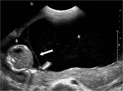
Figure 20.4 Polyhydramnios-oligohydramnios sequence in twin-to- twin transfusion syndrome.
Table 20.1 Quintero staging of TTTS
| Stage | Description |
| I | Discrepancy in amniotic fluid volume with oligohydramnios of a maximum pool depth (MPD) ≤2 cm in one sac and polyhydramnios in other sac (MPD ≥8 cm). The bladder of the donor twin is visible and Doppler studies are normal |
| II | The bladder of the donor twin is not visible (during length of examination, usually around 1 hour) but Doppler studies are not critically abnormal |
| III | Doppler studies are critically abnormal in either twin and are characterized as abnormal or reversed end-diastolic velocities in the umbilical artery, reverse flow in the ductus venosus or pulsatile umbilical venous flow in either twin |
| IV | Ascites, pericardial or pleural effusion, scalp oedema, or overt hydrops present in recipient twin |
| V | One or both babies are dead |
Source data from Quintero RA, Morales WJ, Allen MH, Bornick PW, Johnson PK, Kruger M. Staging of twin-twin transfusion syndrome.J Perinatol 1999;19:550-5.
there should be detailed ultrasound assessment and testing for karyotype abnormalities (19).
The gold standard treatment for TTTS diagnosed before 26 weeks is laser ablation, as the evidence suggests that it leads to better outcomes compared with amnioreduction or septostomy (48). Indeed, the Eurofetus randomized trial showed a higher survival after fetoscopic laser (76% of at least one twin) compared with serial amnioreduction (56%) (49). However, the Cochrane review on interventions in twin-to-twin transfusion also suggested that if laser ablation expertise is not available, amnioreduction is an acceptable alternative in pregnancies diagnosed after 26 weeks of gestation. Techniques described in fetoscopic laser for TTTS include the non- selective technique for ablation of all the vessels that cross the intertwin membrane, the selective technique which identifies specific anastomosis and ablates them, and lastly the ‘connecting the dots' or Solomon technique, which involves lasering healthy areas of the placenta between lasered anastomoses in order to create an ‘equator' or ‘dichorionization' of placenta (50-52).
Following laser treatment, the recurrence rate of TTTS is up to 14%, and this risk is reduced by use of the Solomon technique compared with the highly selective technique (53-55).Fetoscopic laser technique is therefore offered for stage II to stage IV TTTS. At present, there is a variation in offering laser versus expectant management for stage I TTTS. In a systematic review of the management of stage I TTTS pregnancy, overall survival appeared to be similar for those undergoing laser therapy or conservative management (85% and 86%, respectively), but was somewhat lower for those undergoing amnioreduction (77%) (56). Indeed, the management option for stage I TTTS will be debatable till the results of a randomized control trial comparing expectant versus laser management are available (NCT01220011) (57). Another option for the management of severe TTTS is selective termination of pregnancy using bipolar diathermy, laser coagulation, or radiofrequency ablation of one of the umbilical cords. Rarely, parents may opt for termination of the entire pregnancy.
From October 2015, the Twins and Multiple Births Association (TAMBA) have supported a United Kingdom-based registry of complications of monochorionic twins with emphasis on TTTS. This will provide a tool to assist the improvement of clinical skills and practice and also establish a platform to allow long-term followup of TTTS survivors at a national level showing the longer-term neurodevelopment outcomes (58).
TTTS has an effect on the cardiovascular status of both babies— possibly more in the recipient. Even early in stage I, up to 55% of fetuses have a degree of myocardial dysfunction (59). Studies have also shown evolution of pulmonary stenosis and functional pulmonary atresia in the recipient twin after laser procedure, suggesting that the cardiovascular status of twins in a pregnancy complicated by TTTS should be evaluated postnatally (60).
In pregnancies complicated by TTTS, cerebral abnormalities have been reported in 5% of those undergoing laser coagulation, 14% following serial amnioreduction, and 21% following expectant management (61). Both donors and recipients are at risk of developing either ischaemic or haemorrhagic lesions. The neurodevelopmental outcome at 6 years of age was similar to that at the age of 2 years and 10 months, with 9% of the children experiencing major neurodevelopmental delay (62). Neurodevelopment outcome was no different in those with TTTS treated by the Solomon technique versus the standard technique in a subgroup analysis of the survivors recruited in the Solomon randomized trial (63).
Selective fetal growth restriction
sIUGR by definition is applicable to cases in monochorionic pregnancies where the estimated fetal weight (EFW) of the small fetus falls below the tenth percentile and the inter-twin growth discordance is greater than 25%. This condition is present in about 10-15% of all monochorionic twin pregnancies and in a small proportion can coexist with a superimposed TTTS. The pathophysiology behind sIUGR is inadequate placental sharing, possibly in association with very eccentric or velamentous cord insertion, and the presence of vascular anastomoses in the monochorionic placenta (64).
sIUGR is divided into three subtypes depending on whether there is umbilical artery Doppler waveform abnormality. Perinatal outcome differs in these three subtypes. In type I sIUGR, the lowest risk type, there is positive end-diastolic flow in the umbilical artery waveform of the growth-restricted fetus, and if this persists, the outcome can be good with over 90% survival. In type II sIUGR, there is persistent absent or reversed end-diastolic flow in the umbilical artery waveform of the growth-restricted fetus, and there is a risk of fetal death of either twin in up to 29% and neurological sequelae in 15% of cases born before 30 weeks. In type III sIUGR, there is a cyclical pattern present—absent and reversed end-diastolic flow in the umbilical artery waveform of the growth- restricted fetus and a 10-20% risk of sudden death of the growth- restricted fetus, and a high rate of neurological morbidity (up to 20%) in the larger twin (65).
There is limited evidence to guide the management of monochorionic twins affected by sFGR. Options include conservative management followed by early delivery, fetoscopic laser ablation, or cord occlusion of the growth-restricted twin (in order to protect the co-twin) (66). Irrespective of the presence or absence of intervention, such pregnancies are at a high risk of iatrogenic preterm delivery (3).
Twin anaemia polycythaemia sequence
The prenatal diagnosis ofTAPS is based on the finding ofa discordant middle cerebral artery (MCA) Doppler—MCA peak systolic velocity (PSV) greater than 1.5 multiples of the median (MoM) in the donor, suggesting fetal anaemia, and MCA PSV less than 1.0 MoM in the recipient, suggesting polycythemia. The incidence of TAPS in MCDA twins is up to 5% (spontaneous) and up to 13% (iatrogenic following laser ablation for TTTS) (67). TAPS occurs due to the presence of small arteriovenous anastomoses (conceptions matched for age and parity (odds ratio: 1.57) (82).
The cause of the high incidence of spontaneous preterm birth in multiple pregnancies is generally attributed to uterine overdistension, precipitating increased myometrial contractility. However, the aetiology is probably more complex. For example, social stress is an important predictor of preterm birth in twin pregnancies (83).
Several factors/tests have been studied as predictors of spontaneous preterm birth in twin and triplet pregnancies, including ultrasonographic cervical length (CL) measurements (one off or serial), fetal fibronectin test, other biomarkers, home uterine activity monitoring, previous history of preterm birth, and a combination of these approaches.
A systematic review of 21 studies comprising 3523 twin pregnancies concluded that transvaginal CL at 20-24 weeks' gestation is a good predictor of spontaneous preterm birth in asymptomatic women with twin pregnancies (84). The NICE guideline recommended that a CL of less than 25 mm at 18-24 weeks' gestation is a good predictor of spontaneous preterm delivery in twin pregnancy and a CL measurement of less than 25 mm at 14-20 weeks' gestation is a good predictor of spontaneous preterm birth in triplet pregnancy (11). A recent study reported serial CL measurements to have a better detection rate (69% vs 28%; P for IUGR or birthweight differences in twin and triplet pregnancies (11). The American College of Obstetricians and Gynecologists considers a difference of 15-25% in the EFW to constitute discordant fetal growth. Based on a consensus of a group of experts in multiple pregnancy, the ISUOG has recently recommended an arbitrary cut-off of 20% for distinguishing pregnancies at increased risk of adverse outcome (19). It must, however, be acknowledged that the discordance cut-off most predictive of adverse outcome is likely to vary with gestational age (99). A United Kingdom-based large cohort study of 2161 twin pregnancies (302 monochorionic and 1859 dichorionic twin pregnancies) showed that both EFW and birthweight discordance are good predictors of adverse outcome (99). The RCOG recommends that growth discordance should be measured in all monochorionic twin pregnancies at 2-weekly intervals from 16 weeks' gestation onwards and an appropriate referral should be made to the regional fetal medicine unit with expertise in multiple pregnancy if the discordance is 20% or more (3).