Infections of the Genital Tract
Sangini Sheth
Jean M. Keller
INFECTIONS OF THE LOWER GENITAL TRACT
Symptoms caused by infections of the lower genital tract are among the most common presenting complaints of gynecologic patients.
This chapter reviews the following: vulvar infections, parasitic infections, ulcerative lesions, vaginitis, cervicitis, and pelvic inflammatory disease (PID). Urinary tract infections are covered in Chapter 16.Vulvar Infections
Human Papillomavirus
• Human papillomavirus (HPV) infection is the most common sexually transmitted disease (STD) in the United States, with an estimated 80% of sexually active women having acquired genital HPV by the age of 50 years. Most HPV infection is asymptomatic or subclinical, with the majority of patients clearing the infection within 2 years.
• There are over 100 types of HPV, of which approximately 30 are mucosal and can infect the lower genital tract in women. HPV types 6 and 11 cause condyloma acuminata or genital warts. HPV can be classified as low-, intermediate-, and highrisk for the development of squamous cell carcinoma, with the majority of cervical cancer caused by HPV types 16 and 18 (see Chapter 45).
• HPV prevalence is highest among 20- to 24-year-olds. Risk factors for HPV infection include number of sexual partners, history of other sexually transmitted infections (STIs), smoking, and immune deficiency such as HIV or use of immunosuppressive medications.
• Signs and symptoms of genital warts include soft, sessile, and/or verrucous lesions on any mucosal or dermal surface that range in size and formation. Lesions are
P.357 usually multifocal and asymptomatic, although itching, burning, bleeding, vaginal discharge, and pain can occur.
• Diagnosis: Genital warts are usually diagnosed by gross inspection, and colposcopic examination may aid in the identification of cervical or vaginal lesions.
HPV testing is not warranted for the diagnosis of genital warts and results would not alter management. Biopsy is recommended if there is no response to standard therapy; the lesions worsen with therapy; there are hyperpigmented, indurated, fixed, ulcerated, bleeding, or atypical lesions; if the patient is immunocompromised; or if the diagnosis is uncertain. Condyloma acuminatum must be differentiated from the lesion of secondary syphilis, condyloma lata.• Treatment: Treatment is indicated for cosmetic benefit and to address symptoms. There are multiple modalities for the treatment of genital warts, including surgical excision, application of topical cytotoxic or keratolytic agents, cytodestructive techniques, and immune modulators. No single treatment method has been shown to be optimal and therefore treatment should be based on patient preference and provider experience. Clinical factors to consider in choosing a treatment modality include anatomic location, size, morphology, and number of lesions. Additional factors that might influence treatment choice include cost of treatment, convenience, and side effects. Lesions may spontaneously regress and recur. A combination of approaches may be required. No therapy can ensure complete eradication of the virus and it remains unclear whether treatment reduces further transmission (T able 28-1). Most lesions resolve within 3 months of treatment; however, recurrence rates range from 30% to 70%.
• Complications from treatment include hypo- or hyperpigmentation of treated areas with ablative or immune- modulating modalities. Rarely, abnormal scarring or chronic pain can occur.
• Prevention: Two types of HPV vaccine are licensed by the U.S. Food and Drug Administration (FDA). Cervarix is a bivalent vaccine that protects against HPV types 16 and 18, which accounts for 70% of all cervical cancer. Gardasil is a quadrivalent vaccine against types 16 and 18, as well as types 6 and 11 found in 90% of genital warts. The HPV vaccine is recommended for female patients 9 to 26 years old and male patients 9 to 21 years old but can be given up to 26 years of age.
Vaccination does not replace routine cervical cancer screening. See also Chapters 45 and 46.Molluscum Contagiosum
• Molluscum is a benign poxvirus infection of the skin found worldwide but is most common in the developing world. It is spread by skin contact (sexual or nonsexual), autoinoculation, and fomites. The incubation period ranges from several weeks to months.
• Signs and symptoms include the appearance of dome-shaped papules with central umbilication ranging from 2 to 5 mm in diameter. Multiple lesions may arise but generally are fewer than 20. The lesions are usually asymptomatic but occasionally pruritic and may become inflamed and swollen. They are usually self-limited, lasting for 6 to 12 months, but may take as long as 4 years to resolve.
• Diagnosis: The characteristic appearance of molluscum contagiosum lends itself to clinical diagnosis by gross inspection. When in doubt, a crush preparation (i.e., microscopic examination of white, waxy material expressed from a nodule) can be performed. Intracytoplasmic eosinophilic inclusion bodies (molluscum bodies) inside keratinocytes confirm the diagnosis. Immunocompromised patients with HIV/AIDS or other conditions can develop giant lesions (>15 mm in diameter) and large numbers of lesions that may be resistant to standard therapy.
P.358
P.359
TABLE 28-1 Treatment Options for Genital Warts
| Therapy | Application | Clearance Rate (%) | Recurrence Rate (%) | Use in Pregnancy | ||||
| Patient applied | ||||||||
| Imiquimod 5% cream | Apply three times a week at bedtime for up to 16 wk. Wash the area with soap and water 6-10 hr after application. | 40-77 | 5-19 | Contraindicated | ||||
| Podofilox 0.5% solution or gel | Apply bid for 3 d, no treatment for 4 d; repeat the cycle for up to four times. Do not exceed 10 | 68-88 | 16-34 | Contraindicated | ||||
| Sinecatechins 15% ointment | cm2 area of treatment or 0.5 mL volume of podofilox per day. Apply three times daily (0.5 cm strand to each wart) until complete resolution of warts (do not exceed 16 wk of treatment). Do not use in immunosuppressed patients or those with clinical genital herpes or open wounds. | 54-57 | 10 | Contraindicated | ||||
| Provider administered | ||||||||
| Podophyllin resin at 10%- 25% in benzoin | Can be repeated one or two times weekly, as needed | 38-79 | 21-65 | Contraindicated | ||||
| 5-Fluorouracil epinephrine gel | Intralesion injection weekly for up to 6 wk | 61 | 50-60 | |||||
| Interferons | Inject at the edge of and beneath the wart with a 26- to 32-gauge needle. | 36-53 | 21-25 | Not recommended | ||||
| Topical trichloroacetic acid or bichloroacetic acid (80%- 90% solution) | Apply small amount one or two times weekly until the wart sloughs off. Typical course is six treatments. | 81 | 36 | Permitted | ||||
| Excisional procedures | Electrocautery or sharp excision may be employed. | 89-93 | 19-22 | Only if obstructing vaginal delivery | ||||
| Cryotherapy with liquid nitrogen | Can be repeated one or two times weekly until resolved | 70-96 | 25-39 | Permitted | ||||
| CO2 laser excision | 72-97 | 6-49 | Not recommended | |||||
bid, twice a day.
From Workowski KA, Berman S; Centers for Disease Control and Prevention. Sexually transmitted diseases treatment guidelines, 2010. MMWR Recomm Rep 2010;59(RR-12):1-110, with permission.
P.360
• Treatment: Molluscum contagiosum is usually self-limited. Multiple regimens have been evaluated in clinical trials, with none being convincingly efficacious. Many practitioners employ watchful waiting. Treatment should be considered, however, in immunosuppressed individuals and those with sexually transmitted lesions that risk infecting their partners. Lesion visibility and patient preference may prompt therapy, which consists of evacuation of the core material with cryofreezing, curettage, or laser ablation.
Parasites
Pediculosis Pubis
• An ectoparasite, Phthirus pubis is usually restricted to the pubic, perineal, and perianal areas but may infect the eyelids and other body parts. It can be transmitted sexually or via close contact through shared bedding or clothing. The parasite deposits eggs at the base of the hair follicle. The incubation period is 1 week and the crab louse lives for about 6 weeks but dies within 24 hours without blood.
• Symptoms of infection include intense pruritus in the affected area, sometimes accompanied by maculopapular lesions. Occurrence of a large number of bites over a short period may lead to systemic manifestations, such as mild fever, malaise, or irritability.
• Diagnosis is made by gross visualization of lice, larvae, or nits in the pubic hair or microscopic identification of crablike lice under oil.
Scabies
• Scabies is caused by the mite Sarcoptes scabiei var. hominis. It is transmitted via prolonged close contact (sexual or nonsexual) and may infect any part of the body, especially flexural surfaces of the elbows, wrists, finger webs, axilla, genitals, and buttocks.
• Fomite transmission is considered possible through clothing, bedding, or towels. The adult female burrows beneath the skin, lays eggs, and travels quickly across the skin.
Crusted or Norwegian scabies is highly infectious and is an aggressive infestation in immunodeficient, debilitated, or malnourished persons, and is associated with increased treatment failure.• Symptoms include the insidious onset of severe intermittent pruritus approximately 3 to 6 weeks after the initial exposure. Subsequent infections can become symptomatic within 24 hours of reinfection. The intense pruritus may worsen at night and include most of the body. The characteristic lesion is the burrow, a 1- to 10mm curving track that serves to house the mite. Other lesions include papules and vesicles.
• Diagnosis can often be made clinically based on history and gross appearance of the burrows. Skin scrapings can be obtained for microscopic examination under oil.
• Treatment (Table 28-2) for pediculosis pubis and scabies requires an agent that kills both adult organisms and eggs. T reatment should include decontamination of clothing and bed linens with dry cleaning or machine washing and drying with hot cycle. Treat pruritus with antihistamines.
• Toxic effects of lindane include seizures and aplastic anemia. This agent is not recommended for use in pregnant or lactating women, children younger than age 2 years, or patients with extensive dermatitis.
P.361
| TABLE 28-2 Treatment Options for Parasites | ||
| Pediculosis Pubis | Scabies | |
| Permethrin (Nix) cream—safe in pregnancy | Apply 1% cream rinse to the affected areas, wash off after 10 min, and comb the infested areas with a finetoothed comb. | Apply 5% cream to all areas of the body from the neck down and wash off after 8-14 hr. |
| Pyrethrins with piperonyl butoxide — safe in pregnancy | Apply to the affected area and wash off after 10 min. | — |
| Ivermectin | 250 pg/kg orally, repeated in 2 wk | 200 pg/kg orally, repeated in 2 wk |
| Malathion | 0.5% lotion applied for 8-12 hr and washed off | — |
| Lindane 1% (Kwell) lotion, cream, or shampoo—not for use in pregnancy | bgcolor=white>—Not first line due to toxicity. Apply 1 oz of lotion or 30 g of cream in a thin layer to all areas of the body from the neck down and thoroughly wash off after 8 hr. | |
| • Treat pruritus with antihistamines. | ||
| • Clothes and linens should be laundered in hot water and heat dried or removed from body contact for at least 72 hr. | ||
| • Sexual partners should be treated. Infected individuals should be tested for other sexually transmitted infections. | ||
| From Workowski KA, Berman S; Centers for Disease Control and Prevention. Sexually transmitted diseases treatment guidelines, 2010. MMWR Recomm Rep 2010;59(RR-12):1-110, with permission. | ||
Genital Ulcers
• The most common infectious causes of genital ulcers in young, sexually active women are herpes simplex virus (HSV) and Treponema pallidum (syphilis). Less common causes include chancroid and donovanosis. Genital herpes is the most prevalent. All of these lesions are associated with increased risk of HIV acquisition. A patient presenting with genital ulcers should be evaluated for syphilis, herpes, and Haemophilus ducreyi in areas where chancroid is prevalent, as well as HIV if status is unknown.
Genital Herpes
• At least 50 million people in the United States have HSV-2 genital herpes, a chronic STI. Multiple types of herpesvirus have been identified. Historically, HSV-2 accounts for the majority of genital infections; however, HSV-1 now accounts for up to 50% of first-episode cases. HSV-1 genital infections are less likely to recur
P.362 and less commonly result in asymptomatic viral shedding. The majority of persons infected with HSV-2 remain undiagnosed and intermittent viral shedding accounts for most HSV transmission.
• Clinical diagnosis of genital herpes is both insensitive and nonspecific. The classic presentation of multiple, painful, vesicular, or ulcerative lesions is absent in many patients. Herpetic outbreaks can last as long as 2 to 6 weeks in a first-episode primary infection and up to 7 days in recurrent outbreaks. Classically, lesions are preceded by vulvar paresthesias or pruritus, followed by the formation of multiple vesicles that coalesce into ulcerations, which may be painful. Outbreaks are selflimiting, and lesions heal without scar formation. The prodrome of itching or burning in the affected area is important for counseling patients on when to start antiviral therapy because systemic symptoms are usually absent. The majority of patients with HSV-2 will experience recurrent outbreaks in the first year with declining frequency over time. Patients should be counseled that asymptomatic shedding of virus with possible transmission to sexual partners can occur in the absence of outbreaks.
• Diagnosis: Clinical suspicion is based on history and the appearance of lesions. Obtain laboratory confirmation with type-specific virologic and serologic testing. Documentation of HSV-1 or HSV-2 is useful for prognosis and counseling.
• Virology: Cell culture and polymerase chain reaction (PCR) testing are preferred for diagnosis of HSV. Sensitivity of cell culture is low, especially with recurrent lesions or those that have begun to heal. PCR is becoming more common due to its increased sensitivity. Viral culture isolates should be typed to determine HSV-1 or HSV-2; lack of HSV detection by culture or PCR does not prove absent infection because viral shedding is intermittent.
• Serology can confirm clinical suspicion in the absence of a positive culture; antibodies develop within weeks of infection. Type-specific assays that differentiate glycoprotein 1 (HSV-1) from glycoprotein 2 (HSV-2) are recommended. The presence of HSV-2 antibodies is predominantly seen in genital infections and patients should therefore be counseled as such. HSV-1 antibodies, however, may be the result of childhood transmission although genital transmission is growing.
• Treatment (Table 28-3)
• Systemic antiviral therapy for HSV may reduce symptoms and complications of infection. Medical management does not eradicate the virus or reduce the frequency or severity of recurrences after medication is stopped. Primary genital herpes outbreaks should be treated with antiviral therapy, as patients are at increased risk for severe or prolonged symptoms.
• Episodic treatment for recurrent herpes should be initiated within 1 day of lesions or during prodromal period.
• Suppressive therapy can reduce recurrence in up to 80% of patients. Daily suppressive therapy with valacyclovir 500 mg a day has been shown to decrease HSV-2 transmission in discordant, heterosexual couples.
• Recurrences will decrease over time regardless of suppressive therapy, so providers should address continued suppressive therapy yearly.
• Severe or complicated disease should be treated with intravenous acyclovir (5 to 10 mg/kg every 8 hours for 2 to 7 days or until clinical improvement is observed followed by oral therapy to complete 10-day course).
• Topical antiviral therapy has not shown any benefit and is not recommended.
• The virus cannot be completely eradicated and remains latent in the cell bodies of sacral nerves S2, S3, and S4.
P.363
TABLE 28-3 Treatment for Genital Herpes
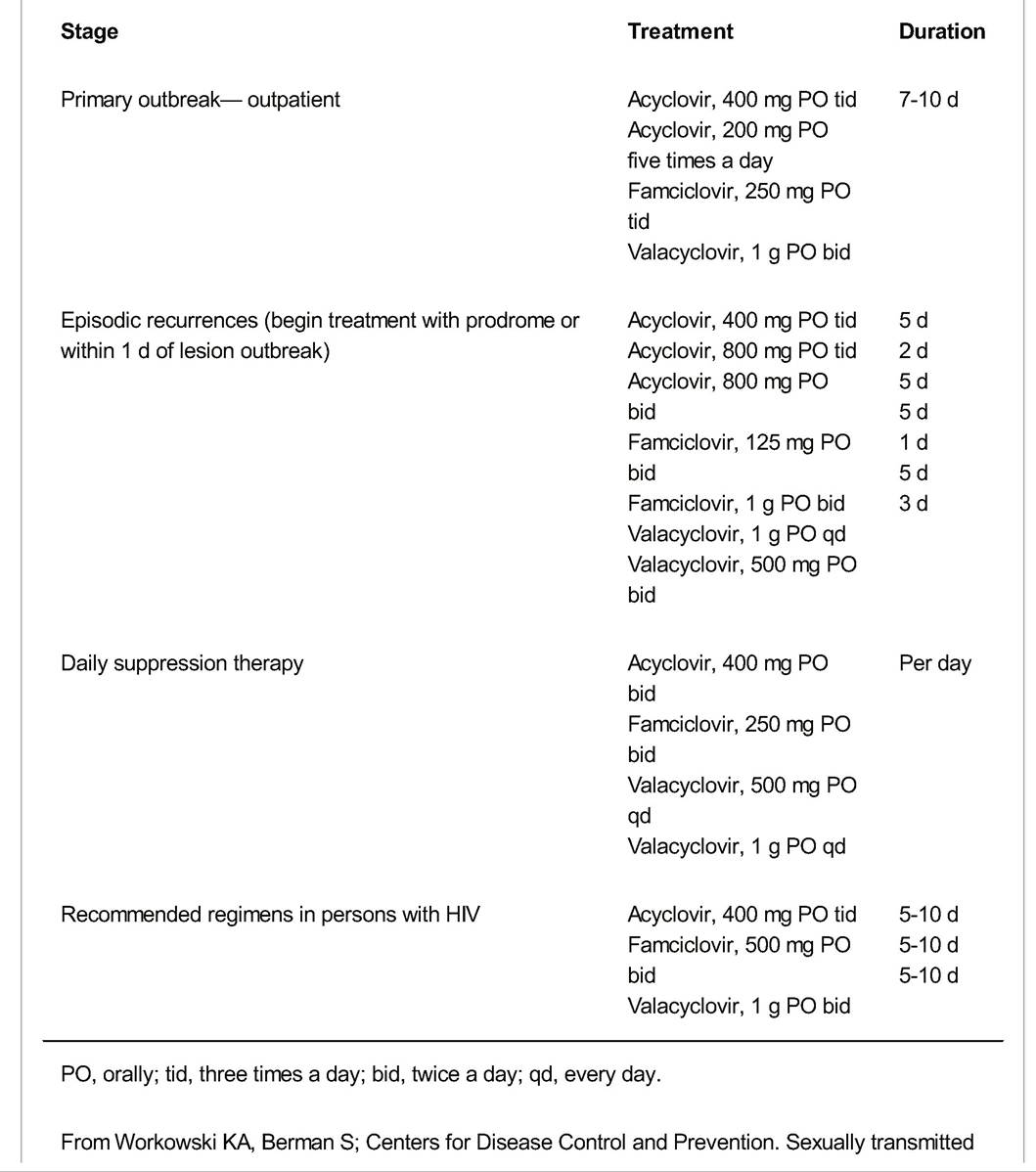
diseases treatment guidelines, 2010. MMWR Recomm Rep 2010;59(RR-12): 1-110, with permission.
• An effective HSV vaccine is not yet available.
• All women with genital herpes should be counseled on the natural history of HSV, sexual and perinatal transmission risks, and ways to reduce transmission.
• Complications include herpes encephalitis (rare but potentially life-threatening) and urinary tract infection (which can cause urinary retention or severe pain).
• Counseling: Patients should be advised to remain abstinent from the onset of prodromal symptoms until complete reepithelialization of lesions. Couples should discuss the role of suppressive therapy in decreasing transmission risk. Counseling should be appropriate to the HSV type.
• During pregnancy, women with primary HSV should be treated with antiviral therapy. Perinatal transmission is possible, and therefore, cesarean delivery is recommended for women with active lesions or prodromal symptoms of genital HSV at delivery. The risk of perinatal HSV transmission is high among women who acquire HSV near time of delivery and low for those with recurrent herpes. Many providers prescribe suppressive therapy for pregnant women with a history of genital herpes beginning at 36 weeks' gestation. Also see Chapter 11.
Syphilis
• The spirochete T. pallidum causes the systemic disease syphilis. The disease is contagious only when mucocutaneous lesions are present. This occurs through contact with a chancre, condyloma lata, or mucosal lesion. The organism can penetrate skin
P.364 or mucous membranes, incubating over a period of 10 days to 3 months. Syphilis has a complex course characterized by the immunologic response to the spirochete.
• Syphilis is divided into overlapping stages: primary, secondary, neurologic, and tertiary based on clinical findings to guide treatment and follow-up. Latent infections without clinical manifestations is detected with serologic testing and characterized as early latent, infection acquired in the previous year or late latent, and infection of greater than 1 year duration or latent syphilis of unknown duration. Determination of early latent versus late latent or latent infection of unknown duration guides duration of therapy.
• Primary syphilis usually presents as a hard, painless, solitary chancre appearing on the vulva, vagina, or cervix, although extragenital lesions may occur. Lesions that occur on the cervix or in the vagina often go unrecognized. Nontender inguinal lymphadenopathy is frequently present. The primary chancre resolves spontaneously within 2 to 6 weeks.
• Secondary syphilis occurs after hematogenous spread of the spirochete and is characterized by protean manifestations including generalized nonpruritic papulosquamous rash typically on the palms and soles, irregular rash, mucous patches, patchy alopecia, condyloma lata, and generalized lymphadenopathy. Systemic symptoms such as fever, headache, and malaise also occur.
• Latent syphilis is defined by seropositivity without evidence of clinical manifestations. Latent syphilis documented as acquired during the previous year is referred to as early latent. All other latent syphilis is either late latent or latent syphilis of unknown duration. The late latent phase (>1 year) is not infectious by sexual transmission, but the spirochete may transplacentally infect the fetus.
• Tertiary syphilis develops in up to one third of the untreated or inadequately treated patients and refers to gummas, locally destructive lesions of the bone, skin, or other organs. Cardiovascular involvement in tertiary syphilis includes aortic aneurysm and aortic valvular insufficiency.
• Neurosyphilis can occur during any stage of syphilis and is not synonymous with tertiary syphilis. All patients with clinical evidence of central nervous system involvement, evidence of active tertiary syphilis, or serologic treatment failure should have examination of the cerebrospinal fluid (CSF) performed. CSF should be tested for fluorescent treponemal antibody absorption (FTA-ABS) reactivity.
• Diagnosis: T. pallidum cannot be cultured in vitro. The diagnosis is made definitively by identifying the spirochete through dark-field microscopy or by direct fluorescent antibody tests of lesion exudate or tissue. The majority of syphilis infection is diagnosed presumptively with nontreponemal serologic tests, such as the Venereal Disease Research Laboratory (VDRL) or rapid plasma reagin (RPR) and treponemal tests. A positive VDRL or RPR requires confirmation with treponemal testing. These are FTA-ABS, T. pallidum passive particle agglutination assay (TP-PA assay), various enzyme immunoassays, and chemiluminescence immunoassays. False-positive nontreponemal tests are associated with pregnancy, autoimmune disorders, chronic active hepatitis, intravenous drug use, febrile illness, and immunization. Serologic tests become positive 4 to 6 weeks after exposure, usually 1 to 2 weeks after the appearance of the primary chancres. The specific FTA-ABS test remains positive indefinitely. Some laboratories have begun screening with treponemal tests which will be positive in individuals with previously treated syphilis as well as those with untreated or incompletely treated syphilis. A positive result must be followed by a nontreponemal test with titer. If the nontreponemal test is negative,
P.365 a different treponemal test should be performed to verify the results of the first test. If the second treponemal test is positive, those without a history of prior treatment should be offered treatment.
• The diagnosis of neurosyphilis cannot be made with a single test but requires a combination of reactive serologic tests, CSF analysis, and reactive VDRL-CSF with or without clinical symptoms.
• The diagnosis of syphilis should prompt HIV testing and if negative repeated again in 3 months for those living in high HIV-risk areas (prevalence >1% of the population).
• Pregnancy: All women should be screened for syphilis in early pregnancy, and this is mandated in most states. In high-risk patients or in high-prevalence areas, syphilis testing should be repeated twice in the third trimester (i.e., at 28 to 32 weeks' gestation and again at delivery).
• Treatment options are listed in Table 28-4. Penicillin G is the recommended treatment for all stages of infection; however, the choice of preparation should be based on stage and clinical symptoms of disease. Individuals with an allergy to penicillin may be desensitized and treated with benzathine penicillin. Intravenous penicillin G is the only treatment with documented efficacy in pregnancy.
• Follow-up: Definitive criteria for treatment cure or failure have not been established. Clinical follow-up and serologic VDRL or RPR titers should be obtained (preferably at same lab) every 6 months for 1 year or at 3, 6, 9, 12, and 24 months if HIV-positive. If signs or symptoms persist, or there is a fourfold increase in titer, then treatment has failed or the patient has been reinfected. If initial high titer >1:32 remains stable or does not decrease fourfold (two dilutions) in 6 months, treatment failure may have occurred. These patients should undergo repeat HIV testing, lumbar puncture for CSF evaluation, and retreatment. For patients with neurosyphilis, if CSF pleocytosis is noted initially, a repeat CSF evaluation should occur every 6 months until the cell count normalizes. If the cell count is not decreasing by 6 months or has not normalized by 2 years, retreatment should be considered.
Other Ulcerative Lesions
• Chancroid is rare in the United States and appears to be declining worldwide as well but may still occur in regions of Africa and the Caribbean. Definitive diagnosis is with detection of H. ducreyi on special culture media not widely available and has a sensitivity of 80%. Probable diagnosis can be made if the following criteria are met: (a) One or more painful genital ulcers are present, (b) no evidence of T. pallidum by dark-field examination or serologic testing 7 days after the onset of ulcers, (c) HSV testing of the ulcer is negative, and (d) the clinical appearance of genital ulcers and regional lymphadenopathy (if present) is typical for chancroid. Successful treatment with antibiotics resolves clinical symptoms but in some cases, scarring can result. Recommended treatment is azithromycin 1 g orally (PO) single dose, ceftriaxone 250 mg intramuscularly single dose, ciprofloxacin 500 mg PO twice a day (bid) (contraindicated in pregnancy) ? 3 days, or erythromycin 500 mg PO three times a day ? 7 days.
• Granuloma inguinale (donovanosis) also occurs rarely in the United States but is endemic in some tropical and developing areas. The genital ulcer is caused by the intracellular bacterium Klebsiella granulomatis. The disease is a slowly progressive ulcerative lesion on the perineum or genitals without regional lymphadenopathy. The lesion is highly vascular and subcutaneous granulomas may occur. The organism is difficult to culture and no FDA-cleared DNA detection tests exist. The diagnosis is made by dark stain and visualization of Donovan bodies. Extragenital infection can occur with extension to the pelvis and dissemination to abdominal organs, bones, or the mouth. Treatment with antimicrobials halt progression of the lesion but relapse can occur 6 to 18 months after effective therapy. Recommended treatment is for 3 weeks or until lesions have completely healed and include doxycycline 100 mg bid ? 3 weeks (recommended), alternatively azithromycin 1 g PO q week ? 3, ciprofloxacin 750 mg PO bid, erythromycin 500 mg four times a day (qid), or trimethoprim-sulfamethoxazole double strength 1 tablet PO bid.
P.366
TABLE 28-4 Recommended Treatment for Syphilis
| Phase | Medication | Dosage | Duration | ||
| Primary, secondary, and early latent syphilis (1 yr) and | Benzathine | colspan=2 bgcolor=white>2.4 million U IM (7.2 millionq wk for | |||
| secondary syphilis without neurosyphilis | penicillin G | U total) | 3 wk | ||
| Penicillin allergy (nonpregnant) | Doxycycline or | 100 mg PO bid | 28 d | ||
| Neurosyphilis | tetracycline Aqueous crystalline penicillin G | 500 mg PO qid 3-4 million U IV q4h (18-24 million U per day) | 28 d 10-14 d | ||
| Alternate regimen (if compliance assured) | Procaine penicillin PLUS probenecid | 2.4 million U IM qd plus 500 mg PO qid | 10-14 d 10-14 d | ||
| Tertiary syphilis (gumma, cardiovascular syphilis) | Benzathine penicillin G | 2.4 million U IM (7.2 million U total) | q wk for 3 wk | ||
| Syphilis during pregnancy | Penicillin Desensitize if allergic | Parenteral regimen appropriate for the stage of syphilis | — | ||
| Primary or secondary syphilis in HIV-positive patients | Benzathine penicillin G Desensitize if allergic | 2.4 million U IM | 1 dose | ||
| Latent syphilis in HIV-positive patients | Benzathine penicillin G Desensitize if allergic | 2.4 million U IM (7.2 million U total) | q wk 3 wk | ||
IM, intramuscular; PO, orally; bid, twice a day; qid, 4 times a day; IV, intravenous; qd, every day.
From Workowski KA, Berman S; Centers for Disease Control and Prevention. Sexually transmitted diseases treatment guidelines, 2010. MMWR Recomm Rep 2010;59(RR-12): 1-110.
P.367
• Lymphogranuloma venereum (LGV) is caused by Chlamydia trachomatis serovars L1, L2, or L3 and presents with tender inguinal and/or femoral lymphadenopathy that is usually unilateral. A self-limited genital ulcer or papule may occur at the site of inoculation. Rectal exposure can result in proctitis or colitis with mucoid hemorrhagic discharge, anal pain, tenesmus, fever, and constipation and if left untreated can result in chronic colorectal fistulas and strictures. Both genital and colorectal lesions can develop secondary bacterial inflectional coinfection including sexually and nonsexually transmitted pathogens.
• Diagnosis is based on clinical suspicion, epidemiologic information, and exclusion of other etiologies with clinical symptoms similar to LGV. Chlamydial testing of genital and lymph node specimens with culture, direct immunofluorescence, or nucleic detection can be used for diagnosis but rectal specimens using nucleic acid amplification tests (NAATs) for C. trachomatis are not FDA cleared. PCR-based genotyping to differentiate LGV from non-LGV C. trachomatis are not widely available. In the absence of specific LGV diagnostics, patients with clinical suspicion of LGV should be treated. Doxycycline for 21 days is the preferred treatment and alternative treatment is erythromycin 500 mg PO qid ? 21days.
Vaginitis
• Vaginitis is characterized by pruritus, discharge, odor, dyspareunia, and dysuria. The most common causes are bacterial vaginosis, vulvovaginal candidiasis, and trichomoniasis. The vagina is normally colonized by several organisms, including Lactobacillus acidophilus, diphtheroids, Candida albicans, Gardnerella vaginalis, Escherichia coli, group B streptococci (GBS), genital Mycoplasmatales and other flora. Its physiologic pH is approximately 4.0, with peroxide-producing L. acidophilus inhibiting overgrowth of pathogenic bacteria. Vaginal fluid is typically white, odorless, and seen in dependent areas of the vagina.
• Diagnosis of vaginitis must begin with a focused history on the constellation of vaginal symptoms, location, duration, relation to menstrual cycle, use of prior treatment, douching, and sexual history. Physical examination should start with inspection of the vulva and include speculum examination to obtain samples for vaginal pH, amine (“whiff”) test, saline wet mount, and potassium hydroxide (KOH) microscopy. DNA amplification tests for Neisseria gonorrhoeae and C. trachomatis may be indicated. The three major types of vaginitis and their distinguishing characteristics are described in Table 28-5.
Bacterial Vaginosis
• Bacterial vaginosis (BV) is the most common cause of vaginitis although most women with BV are asymptomatic. No single infectious agent is responsible; rather, there is a shift in the composition of vaginal flora, with up to a 10-fold increase in facultative anaerobic bacteria such as G. vaginalis, Mycoplasma hominis, Atopobium
P.368 vaginae and Prevotella, Bacteroides, Peptostreptococcus, and Fusobacterium species, with a decrease in the concentration of Lactobacillus species. It has been implicated as a risk factor for preterm premature rupture of membranes and preterm delivery. The microbial alterations associated with BV are not completely understood or whether it is associated with a sexually transmitted pathogen; however, it has been associated with multiple male and female partners, new sex partner, lack of condom use, douching, decrease in vaginal lactobacillus, increased risk of STIs, PID, and postprocedural gynecologic infections. T reatment of BV prior to an abortion or hysterectomy decreases the risk of postoperative infection.
TABLE 28-5 Distinguishing Characteristics of Vaginitis
| Bacterial Vaginosis | Trichomonas Vaginitis | Candidal Vaginitis | |
| Vaginal pH | >4.5 | 5.0-7.0 | — |
| Vaginal secretions | Thin, white, adherent; amine (fishy) odor with potassium hydroxide (KOH) | Thin, frothy, white, gray, yellow; copious | Thick, white, curdlike |
| Wet preparation | Clue cells, few WBCs | Trichomonads, WBCs | Hyphae and buds, WBCs (best seen with KOH prep) |
WBCs, white blood cells.
From Amsel R, Totten PA, Spiegel CA, et al. Nonspecific vaginitis. Diagnostic criteria and microbial and epidemiologic associations. Am J Med 1983;74(1): 14-22.
Diagnosis: BV is diagnosed by the presence of at least three of the Amsel clinical criteria: (a) homogenous thin white discharge coating the vaginal walls, (b) vaginal pH >4.5, (c) more than 20% of epithelial cells appearing to be clue cells on microscopic examination, and (d) fishy odor before or after the addition of 10% KOH to the sample (amine test). Detection of three of these criteria has been correlated to Gram stain, considered the gold standard for BV diagnosis, which determines the concentration of lactobacillus to other organisms. Commercially available point-of-care card tests to detect elevated pH and trimethylamine are now available and may be useful when a microscope is not available.
• Treatment: Treatment regimens are shown in Table 28-6 and are recommended for women with symptoms. Benefits of treatment in nonpregnant include amelioration of the signs and symptoms of BV and reduction in the risk of acquisition of HIV, N. gonorrhoeae, C. trachomatis, and other viral STIs.
• BV has been associated with adverse pregnancy outcomes, including premature rupture of membranes, preterm labor, preterm birth, intra-amniotic infection, and postpartum endometritis, although the only established benefit in pregnant women is the reduction of symptoms. Currently, treatment is recommended for
P.369 symptomatic pregnant women only. T reatment of male partners is not necessary or beneficial in preventing recurrence.
TABLE 28-6 Treatment for Bacterial Vaginosis
| Medication | Dosage | Duration | Use in Pregnancy |
| Metronidazole | 500 mg PO bid | 7 d | Recommended |
| Clindamycin phosphate cream 2% | 1 full applicator (5 g) intravaginally qhs | 7 d | Not recommended |
| Metronidazole gel 0.75% | 1 full applicator (5 g) intravaginally daily | 5 d | Not recommended |
| Alternative regimensa | |||
| Clindamycin ovules | 100 g intravaginally qhs | 3 d | Not recommended |
| Clindamycin hydrochloride | 300 mg PO bid | 7 d | Recommended |
| Tinidazole | 2 g PO daily | 2 d | Not recommended |
| Tinidazole | 1 g PO daily | 5 d | Not recommended |
aExtended-release metronidazole (750 mg) and single-dose clindamycin intravaginal cream are
also available. Metronidazole 250 mg PO tid for 7 days is also recommended in pregnancy.
PO, orally; bid, twice a day; qhs, at bedtime; tid, three times a day.
From Workowski KA, Berman S; Centers for Disease Control and Prevention. Sexually transmitted diseases treatment guidelines, 2010. MMWR Recomm Rep 2010;59(RR-12): 1-110.
• Follow-up: Recurrence of BV is common, recurring in up to 30% of women within 3 months. Either retreatment with the same therapy or a different treatment option is acceptable for early treatment failure. Women with multiple recurrences may benefit from metronidazole gel twice weekly for 4 to 6 months after completing a treatment course. Follow-up in 1 month for asymptomatic pregnant women at high risk for preterm delivery should be considered.
Trichomoniasis
• Trichomoniasis is an STI with 7.4 million cases in the United States annually and is caused by the unicellular protozoan Trichomonas vaginalis. Trichomonads can survive on wet towels and other surfaces and thus can be nonsexually transmitted. Its incubation period ranges from 4 to 28 days.
• Diagnosis: Vaginal exam may reveal a frothy, malodorous yellow-green discharge with vulvar irritation and the cervix may appear erythematous and friable. However, many women have minimal or no symptoms. A wet smear preparation that is promptly reviewed may reveal the flagellated, mobile protozoon with a sensitivity of approximately 60% to 70%. Trichomonas culture tests have 90% sensitivity. Point-of-care tests are available and have higher sensitivity than vaginal examination, but false-positives can occur. Culture of secretions is sensitive and highly specific and should be obtained when microscopic evaluation is negative in women who have clinical suspicion for trichomonas. An FDA-cleared test for gonorrhea and chlamydia has been modified to detect T. vaginalis in vaginal, endocervical, or urine specimens and sensitivity range from 88% to 97% and specificity of 98% to 99% is another diagnostic option. Liquid-based testing Pap tests have increased
P.370 sensitivity for T. vaginalis; however, false positives have occurred and confirmatory testing may be needed.
• Treatment consists of one 2-g dose of either metronidazole or tinidazole PO. Alternatively, metronidazole 500 mg PO bid for 7 days can be used. Metronidazole gel has an efficacy of 2% cream 5 g (sustained release), single application
Clotrimazole 1 % cream 5 g intravaginally for 7-14 da
Clotrimazole 2% cream 5 g intravaginally for 3 da
Clotrimazole 100 mg vaginal tablet for 7 d
Miconazole 2% cream 5 g intravaginally for 7 da
Miconazole 4% cream 5 g intravaginally for 3 da
Miconazole 100 mg vaginal suppository, one suppository for 7 da
Miconazole 200 mg vaginal suppository, one suppository for 3 da
Miconazole 1200 mg vaginal suppository, 1 suppository for 1 d
Nystatin 100,000-unit vaginal tablet, one tablet for 14 d
Tioconazole 6.5% ointment 5 g intravaginally in a single application13
Terconazole 0.4% cream 5 g intravaginally for 7 d
Terconazole 0.8% cream 5 g intravaginally for 3 d
Terconazole 80 mg vaginal suppository, one suppository for 3 d
Oral agent
Fluconazole 150 mg oral tablet, one tablet in single dose
aOver-the-counter (OTC) preparations.
From Workowski KA, Berman S; Centers for Disease Control and Prevention. Sexually transmitted diseases treatment guidelines, 2010. MMWR Recomm Rep 2010;59(RR-12): 1-110.
• Treatment: Symptomatic patients, including pregnant women, should be treated. See Table 28-7. Medications available over-the-counter include butoconazole, clotrimazole, miconazole, and tioconazole. Only topical therapies are recommended in pregnancy. For severe VVC, extended treatment with topical azoles up to 14 days or fluconazole 150 mg in two doses 72 hours apart is recommended. Maintenance therapy with 150 mg of fluconazole or clotrimazole 200 mg twice a week or 500 mg once a week is recommended. Maintenance therapy is effective in reducing recurrence in up to 50% of women.
• Follow-up: If symptoms persist or recur within 2 months, patients should return for follow-up.
• Treatment of male partners is not indicated unless the partner has symptoms of yeast balanitis or in cases of recurrent VVC.
Cervicitis
• Cervicitis is characterized by two major diagnostic signs: a purulent or mucopurulent cervical exudate and/or sustained cervical bleeding in response to manipulation by an examining swab. Patients may be asymptomatic but commonly report abnormal vaginal discharge and intermenstrual bleeding. The primary pathogens of mucopurulent cervicitis are the two sexually transmitted organisms C. trachomatis and N. gonorrhoeae; however, trichomoniasis and genital herpes infections can also be associated with cervicitis. In the majority of cases, no etiologic agent is identified; limited data has implicated BV, Mycoplasma genitalium, and frequent douching as other causes of cervicitis. Leukorrhea (>10 white blood cell per high-power field
P.372
on microscopy) of vaginal fluid may be associated with chlamydial or gonorrheal infections. Patients with symptoms of cervicitis should also be evaluated for PID, C. trachomatis, N. gonorrhoeae, T. vaginalis, and BV.
Chlamydia
Chlamydia trachomatis is the most frequently reported sexually transmitted bacterial disease in the United States. Risk factors include age younger than 25 years old, low socioeconomic status, multiple sex partners, and unmarried status. Sequelae of C. trachomatis infection in women may include PID, ectopic pregnancy, chronic pelvic pain, and infertility.
Microbiology: C. trachomatis is an obligate intracellular organism that preferentially infects the squamocolumnar cells in the transition zone of the cervix.
Signs and symptoms: Chlamydial cervicitis is asymptomatic in about 75% of cases. Patients with C. trachomatis infection may complain of abnormal vaginal discharge, burning with urination, spotting, or postcoital bleeding. A yellow mucopurulent discharge may be present.
Diagnosis: NAAT using PCR is the preferred method of diagnosis for chlamydial and gonorrheal cervicitis and can be performed on vaginal, cervical, or urine samples. Screening programs have shown reduction in the rate of chlamydia infection and incidence of PID; therefore, annual screening is recommended for all sexually active women younger than the age of 25 years and in women older than 25 years who present with risk factors. Women with cervicitis should also be evaluated for trichomoniasis and BV (Table 28-8).
Treatment: Presumptive therapy can be initiated based on clinical findings and STD risk assessment (Table 28-9). T reatment for coinfection with gonorrhea is recommended if local prevalence is >5%. Concomitant treatment for BV or trichomoniasis should be given if detected. Sexual partners should be referred to a clinic for treatment.
| TABLE 28-8 Testing for Gonorrhea and Chlamydia Infection | ||||
| N. gonorrhoeae | C. trachomatis | |||
| Sensitivity | Specificity | Sensitivity | Specificity | |
| Endocervical culture | 70-85 | 100 | 60-70 | 100 |
| Immunoassay | >80 | 97-100 | Not reliable | |
| Nucleic acid probe | 77-97 | 96-100 | 92 | 99.7 |
| PCR/LCR | 95 | 100 | 96.7 | 99.7 |
| Values are in percentages. | ||||
| PCR, polymerase chain reaction; LCR, ligase chain reaction. | ||||
Adapted from Black CM. Current methods of laboratory diagnosis of Chlamydia trachomatis infections. Clin Microbiol Rev 1997;10(1):160-184; Van Dyck E, Ieven M, Patten S, et al. Detection of
Chlamydia trachomatis and Neisseria gonorrhoeae by enzyme immunoassay, culture, and three nucleic acid amplification tests. J Clin Microbiol 2001 ;39(5):1751-1756; Koumans EH, Johnson RE, Knapp JS, et al. Laboratory testing for Neisseria gonorrhoeae by recently introduced nonculture tests: a performance review with clinical and public health considerations. Clin Infect Dis 1998;27: 1171-1180.
P.373
TABLE 28-9 Treatment for Chlamydia trachomatis
| Medication | Dosage | Duration | Use in Pregnancy |
| Recommended | |||
| Azithromycin | 1 g PO | 1 dose | Recommended |
| Doxycycline | 100 mg PO bid | 7 d | Contraindicated |
| Amoxicillin (in pregnant women) | 500 mg PO tid | 7 d | Acceptable regimen for pregnancy only |
| Alternative | |||
| Erythromycin base | 500 mg PO qid | 7 d | Alternative |
| Erythromycin ethylsuccinate | 800 mg PO qid | 7 d | Alternative |
| Ofloxacin | 300 mg PO bid | 7 d | Contraindicated |
| Levofloxacin | bgcolor=white>500 mg PO qd7 d | Contraindicated |
PO, orally; bid, twice a day; tid, three times a day; qid, four times a day; qd, every day.
From Workowski KA, Berman S; Centers for Disease Control and Prevention. Sexually transmitted diseases treatment guidelines, 2010. MMWR Recomm Rep 2010;59(RR-12): 1-110.
• Follow-up: Test of cure is not recommended except in pregnancy (4 to 5 weeks after treatment) unless noncompliance or reinfection is suspected or if symptoms persist. Both patient and partner should sustain from intercourse for 1 week after treatment to avoid reinfection. Retesting 3 months after treatment is recommended in order to assess for reinfection.
Gonorrhea
• Risk factors for gonorrhea are essentially the same as those for C. trachomatis.
• Microbiology: N. gonorrhoeae is a Gram-negative diplococcus that infects columnar or pseudostratified epithelium. Genital, pharyngeal, and disseminated infections may occur. The incubation period is 3 to 5 days.
• Signs and symptoms: In women, symptoms often go unrecognized until complications (PID) have occurred. When present, symptoms include vaginal discharge, dysuria, or abnormal uterine bleeding. The most commonly infected site is the endocervix.
• Diagnosis: Culture, nucleic acid hybridization tests, and NAATs are available for the diagnosis of urogenital N. gonorrhea. Culture and nucleic acid hybridization tests require endocervical specimens, whereas NAATs allows the widest variety of specimen sites including endocervical, vaginal, urethral, and urine. NAATs have greater sensitivity than culture; however, culture is the most widely available option for detection in the nongenital sites and is used when antibiotic sensitivity testing is indicated (see Table 28-8). Targeted screening of women younger than 25 years old or those at risk for infection (multiple sexual partners, prior history of gonorrhea or other STI, inconsistent barrier protection use, those with a history of commercial sex work or drug use, women living in areas of high prevalence) is recommended. All women positive for gonorrhea should be tested for other STIs such as chlamydia, HIV, and syphilis.
P.374
• Treatment options are listed in Table 28-10. Due to increasing resistance, fluoroquinolones are no longer recommended for the treatment of N. gonorrhea; cephalosporins are the only recommended class of antimicrobials. Because coinfection with C. trachomatis is common, treatment of both is recommended unless the NAAT is negative. Sexual partners should be referred for treatment. Although azithromycin 2 g PO is effective against uncomplicated gonorrhea, concerns about resistance development, expense, and gastrointestinal upset should limit its use.
• Follow-up with a test of cure several weeks after treatment for uncomplicated gonorrhea is not required. Retesting 3 months after treatment is recommended in order to assess for reinfection.
TABLE 28-10 Treatment for Neisseria gonorrhoeae
Uncomplicated gonococcal infections of the cervix, urethra, and rectuma
Recommended regimens
Ceftriaxone 250 mg intramuscular (IM) in a single dose
OR
Cefixime 400 mg orally once or 400 mg by suspension (200 mg/5 mL)
OR
Single-dose injectable cephalosporin
PLUS
T reatment for chlamydia unless ruled out
Uncomplicated gonococcal infections of the pharynxa
Recommended regimens
Ceftriaxone 125 mg IM in a single dose
PLUS
Treatment for chlamydia unless ruled out
Disseminated gonococcal infection (DGI)
Recommended regimen
Ceftriaxone 1 g IM or IV every 24 hr
Alternative regimens
Cefotaxime 1 g IV every 8 hr
OR
Ceftizoxime 1 g IV every 8 hr
Oral follow-up regimen
Cefixime 400 mg orally twice daily
OR
Cefixime 400 mg suspension (200 mg/5 mL) twice daily
Any regimen should be continued for 24-48 hr after clinical improvement then switch to oral medicines to complete 1 wk of therapy.
aThese regimens are recommended for all adult and adolescent patients, regardless of travel history or sexual behavior.
IV, intravenous.
From Workowski KA, Berman S; Centers for Disease Control and Prevention. Sexually transmitted
diseases treatment guidelines, 2010. MMWR Recomm Rep 2010;59(RR-12): 1-110.
P.375
INFECTIONS OF THE UPPER GENITAL TRACT
Pelvic Inflammatory Disease
• PID is an inflammatory disorder of any combination of the endometrium, fallopian tubes, ovaries, myometrium, parametrium, and pelvic peritoneum.
• Pathophysiology and microbiology: PID is caused by the spread of infection via the cervix. PID is often associated with STIs such as N. gonorrhoeae and C. trachomatis; however, numerous exogenous and endogenous microorganisms may be involved, including anaerobes, G. vaginalis, Haemophilus influenza, Gramnegative rods, Streptococcus agalactia, and enteric species. Other organisms include Mycoplasma spp., Ureaplasma spp., and cytomegalovirus. Diagnosis of PID should include testing for N. gonorrhea and C. trachomatis and screening for HIV.
• Prevention: There are no signs and symptoms pathognomic for PID, and the clinical picture does not accurately predict the extent of tubal involvement. A high degree of suspicion for PID and treatment based on minimal or subtle signs may help reduce the incidence of long-term sequelae. Episodes of PID may have unrecognized or absent symptoms; delays in diagnosis and treatment contribute to serious sequelae. One in four women with PID will experience tubal infertility, an ectopic pregnancy, or chronic pelvic pain. Treatment of sexual partners and education is important in reducing the rate of recurrent infections.
• Risk factors include a previous history of PID, multiple sex partners, adolescence, BV, and current infection by a sexually transmitted organism. A woman's risk of PID is not increased by intrauterine device (IUD) use. See Chapter 32.
• Signs and symptoms: The most common presenting symptom is abdominopelvic pain. Other symptoms include vaginal discharge, dyspareunia, abnormal bleeding, right upper quadrant pain, fever and chills, nausea, and dysuria.
• Diagnosis of PID is difficult because the presenting signs and symptoms vary widely. Health care providers should maintain a low threshold for the diagnosis of PID based on minimal clinical criteria.
• Minimal criteria: Empiric treatment should be initiated in sexually active young women and other women at risk for STDs if they are experiencing pelvic or lower abdominal pain, if no other cause can be identified, and if one or more of the following are present on pelvic examination:
î Cervical motion tenderness
î Uterine tenderness
î Adnexal tenderness
• Additional criteria for diagnosis, which increase specificity
î Oral temperature >101°F (>38.3°C)
î Abnormal cervical or vaginal mucopurulent discharge
î Predominance of white blood cells on saline microscopy of vaginal secretions
î Elevated erythrocyte sedimentation rate
î Elevated C-reactive protein
î Laboratory documentation of cervical infection with N. gonorrhoeae or C. trachomatis
• PID specific criteria
î Endometrial biopsy with histopathologic evidence of endometritis
î Transvaginal sonography or magnetic resonance imaging techniques showing thickened, fluid-filled tubes with or without free pelvic fluid or tuboovarian complex
î Laparoscopic abnormalities consistent with PID
P.376
• Treatment goals for PID are the prevention of tubal damage that leads to infertility and ectopic pregnancy and prevention of chronic infection. Many patients can be successfully treated as outpatients, and early ambulatory treatment should be the initial therapeutic approach once a presumptive diagnosis has been established. Antibiotic choice should provide broad empiric coverage targeting the major etiologic organisms (N. gonorrhoeae and C. trachomatis) but should also address the polymicrobial nature of the disease (Table 28-11). Negative endocervical screening for N. gonorrhoeae and C. trachomatis does not rule out possibility of upper reproductive tract infection. Patients with tuboovarian abscesses should be admitted for at least 24 hours direct inpatient observation and should receive clindamycin or metronidazole with doxycycline for continued oral therapy after transition from parenteral therapy for adequate anaerobic coverage. Due to quinolone resistance in N. gonorrhoeae, quinolone-based regimens are not recommended except in situations where cephalosporins are not feasible. Among women with an IUD in place, there is not enough evidence to recommend IUD removal with acute PID; however, close clinical follow-up is necessary.
• Criteria for hospitalization
• Surgical emergencies (e.g., appendicitis) cannot be excluded.
• Pregnancy
• Failure to respond clinically to oral antimicrobial therapy
• Inability to follow or tolerate an outpatient oral regimen
• Severe illness, nausea and vomiting, or high fever
• Suspected tuboovarian abscess (at least 24 hours of direct inpatient observation recommended)
• Follow-up: Clinical improvement should be noted within 3 days of initiating treatment; those not improving will typically require hospitalization, diagnostic tests, and possible surgical intervention.
• Sequelae: Approximately 25% of PID patients experience long-term sequelae. Infertility due to tubal occlusion affects anywhere from 6% to 60% of women following an episode of PID, depending on severity, whereas the risk of ectopic pregnancy is approximately 6 to 10 times the normal. Chronic pelvic pain and dyspareunia have also been reported and are associated with the presence of adhesive disease and the number of episodes. Fitz-Hugh-Curtis syndrome is the development of fibrous perihepatic adhesions resulting from the inflammatory process of PID that may result in acute right upper quadrant pain and tenderness.
Endometritis (Nonpuerperal)
• Pathophysiology: Endometritis is caused by the ascension of pathogens from the cervix to the endometrium. Pathogens include C. trachomatis, N. gonorrhoeae, Ureaplasma urealyticum, and M. genitalium. Chronic endometritis is often linked to common bacteria such as streptococci, staphylococci, and E. coli. Organisms that produce BV may also produce histologic endometritis, even in women without symptoms. Endometritis is also an important component of PID and may be an intermediate stage in the spread of infection to the fallopian tubes.
Signs and symptoms:
• With chronic endometritis, many women are asymptomatic. The classic symptom of chronic endometritis is intermenstrual vaginal bleeding. Postcoital bleeding, menorrhagia, and a dull, constant lower abdominal pain are other complaints.
• In acute endometritis, uterine tenderness is common.
P.377
P.378
TABLE 28-11 Treatment for Pelvic Inflammatory Disease
Parenteral treatment for severe PID
Transition to oral therapy, which usually can be initiated within 24-48 hr of clinical improvement, should be guided by clinical experience.
Recommended parenteral regimen A
Cefotetan 2 g IV every 12 hr
OR
Cefoxitin 2 g IV every 6 hr
PLUS
Doxycycline 100 mg orally or IV every 12 hr (IV and oral administration have similar bioavailability; administer orally when possible)
Recommended parenteral regimen B
Clindamycin 900 mg IV every 8 hr
PLUS
Gentamicin loading dose IV or IM (2 mg/kg body weight) followed by maintenance dose (1.5 mg/kg) every 8 hr. Single daily dosing (3-5mg/kg) may be substituted.
Alternative parenteral regimen
AmpicillinZsulbactam 3 g IV every 6 hr
PLUS
Doxycycline 100 mg orally or IV every 12 hr
Transition from parenteral to oral regimen
Doxycycline 100 mg PO bid
OR
Clindamycin 450 mg qid
To complete a 14-day treatment course. Clindamycin preferred oral agent in setting of tuboovarian abscess.
Oral treatment for mild to moderate PID
Parenteral and oral therapies have similar efficacy in treatment of women with mild to moderate PID. Women who do not respond to oral therapy within 72 hr should be reevaluated to confirm the diagnosis and should be administered parenteral therapy on either an outpatient or inpatient basis.
Recommended oral regimen
Ceftriaxone 250 mg IM in a single dose
PLUS
Doxycycline 100 mg orally twice a day for 14 d
WITH OR WITHOUT
Metronidazole 500 mg orally twice a day for 14 d
OR
Cefoxitin 2 g IM in a single dose and
Probenecid 1 g orally administered concurrently in a single dose
PLUS
Doxycycline 100 mg orally twice a day for 14 d
WITH OR WITHOUT
Metronidazole 500 mg orally twice a day for 14 d
OR
Other parenteral third-generation cephalosporin (e.g., ceftizoxime or cefotaxime)
PLUS
Doxycycline 100 mg orally twice a day for 14 d
WITH OR WITHOUT
Metronidazole 500 mg orally twice a day for 14 d
For full details and alternative oral treatment regimens, see updated Centers for Disease Control and Prevention guidelines.
IV, intravenous; IM, intramuscular; PO, orally; bid, twice a day; qid, four times a day.
Adapted from Workowski KA, Berman S; Centers for Disease Control and Prevention. Sexually transmitted diseases treatment guidelines, 2010. MMWR Recomm Rep 2010;59(RR-12):1-110.
• Diagnosis: The diagnosis of chronic endometritis is established by endometrial biopsy and culture. The classic histologic findings of chronic endometritis are an inflammatory reaction of monocytes and plasma cells in the endometrial stroma (five plasma cells per high-power field). A diffuse pattern of inflammatory infiltrates of lymphocytes and plasma cells throughout the endometrial stroma or even stromal necrosis is associated with severe cases of endometritis.
• Treatment: The treatment of choice for chronic endometritis is doxycycline, 100 mg PO bid for 10 days. Broader coverage of anaerobic organisms may also be considered, especially in the presence of BV. When endometritis is associated with acute PID, the treatment should focus on the major etiologic organisms, including N. gonorrhoeae and C. trachomatis, and should also include broader polymicrobial coverage.
• See Chapter 23 for puerperal endomyometritis.
SUGGESTED READINGS
American College of Obstetricians and Gynecologists. ACOG practice bulletin no. 57: gynecologic herpes simplex virus infections. Obstet Gynecol 2004;104:1111-1117.
American College of Obstetricians and Gynecologists. ACOG practice bulletin no. 72: vaginitis. Obstet Gynecol 2006; 107:1195-1206.
Biggs WS, Williams RM. Common gynecologic infections. Prim Care Clin Office Pract 2009; 36:33-51.
Leone PA. Scabies and pediculosis pubis: an update of treatment regimens and general review. Clin Infect Dis 2007;44:S153-S159.
Mers D, Wolff T, Gregory K, et al. USPSTF recommendations for STI screening. Am Fam Physician 2008;77(6):819-824.
Soper DE. Pelvic inflammatory disease. Obstet Gynecol 2010;116:419-428.
Wiley D, Masongsong E. Human papillomavirus: the burden of infection. Obstet Gynecol Surv 2006;61(6):S3-S14.