34 Pediatric Gynecology
Sara Seifert
Dayna Burrel
Pediatric gynecology presents many challenges to the general obstetriciangynecologist unaccustomed to dealing with these young patients. Most of the obstacles may be overcome by communicating effectively and allowing the patient to feel “in control.”
• The interview is the most important aspect in determining the true reason for the visit.
Due to different levels of maturity in each age group of children, different approaches to communication may be used. Including parental figures in the discussion is key.• Gynecologic problems often experienced by the pediatric patient include vulvovaginitis, trauma, foreign bodies, prepubertal vaginal bleeding, abnormal pubertal development, urogenital abnormalities, and genital tumors.
GYNECOLOGIC EVALUATION OF A PREPUBERTAL CHILD
• The examination presents a unique set of difficulties that may be overcome by following a few key guidelines:
• Give the patient a sense of control.
• Display a caring and gentle attitude at all times; the initial evaluation can set the tone for all future examinations.
• The physical exam should include an overall assessment of other organ systems. This allows the patient to feel more comfortable in the exam room and the
P.452 examiner to gain an overall appreciation of height, weight, skin disorders, hygiene, and other indicators of pubertal development.
• If the child is very young or has suffered physical abuse, she may need to be evaluated under anesthesia.
• Make it clear to the child that the examination is permitted by her caregiver and that if anyone else tries to touch her genital area, she should tell her caregiver.
• A chaperone should be present during the physical exam.
General Pediatric Physical Exam
• The abdominal exam can be facilitated by placing the child's hand over the examiner's hand.
• Palpate the inguinal regions to identify potential hernias or gonadal masses.
• Tanner classification of the external genitalia and breast development should be used to quantify pubertal changes (Fig. 34-1).
Pediatric Pelvic Exam: Positioning
• Frog-leg posture: child supine with feet together and knees bent outward. Commonly used in the younger patient.
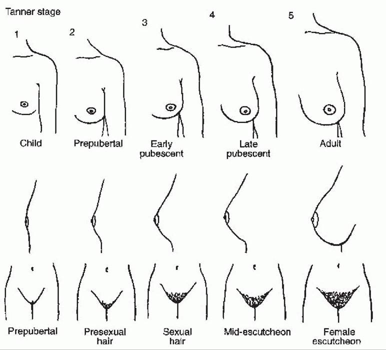
Figure 34-1. Tanner stages of development. (From Beckmann CR, Ling FW, Barzansky BM, et al. Obstetrics and Gynecology, 2nd ed. Baltimore, MD: Lippincott Williams & Wilkins, 1995:8, with permission.)
P.453
• Knee-chest position: when combined with a Valsalva maneuver, allows for assessment of the introital area.
Using an otoscope for magnification or nasal speculum may help with visualization when the primary complaint is vaginal discharge or foreign bodies.
• Supine lateral-spread method: often sufficient enough to allow for visualization of the vestibular structures
• Mother’s lap positioning: Allow the patient to sit in her mother's lap, knees bent, heels on mom's knees; combine with lateral traction of the labia for adequate exposure.
• When a child is uncooperative or evaluation of the genitalia is not optimal, an exam under anesthesia or a return visit may be necessary.
Pediatric Pelvic Exam: Assessment
• Note perineal hygiene, presence of pubic hair, hymenal configuration, size of the clitoris, and the presence of vulvovaginal lesions or vaginal discharge.
• Careful inspection of the hymen must be completed before pelvic examination. A Foley catheter balloon can be placed behind the hymen and filled to visualize a redundant hymen.
• Lateral downward traction of the labia allows visualization of the hymen in prepubertal girls.
• Specimens may be collected using a small urethral Dacron swab. A second technique employs an empty butterfly catheter attached to a syringe and saline is flushed and aspirated to obtain a mix of secretions.
A pediatric feeding tube attached to a 20-mL syringe also allows for vaginal irrigation.• Use of “extinction stimuli” can greatly facilitate a first pelvic exam. Use a distracting stimulus to draw attention from a second stimulus. For example, press a nonexamining finger into the patient's perineum before touching the introitus and allow the patient to acknowledge the presence of its pressure.
• Proper instrument selection is important. Speculum exams are rarely appropriate in the prepubertal patient. Often, the hymenal ring is too tight to accommodate even a pediatric speculum. A nasal speculum can be used for an exam under anesthesia if speculum exam is necessary.
• Rectoabdominal examination may aid in examination of the uterus in a patient who cannot tolerate a vaginal exam.
• Common exam findings:
• Newborn child: It is important to recognize that maternal estrogen influences physical development of the newborn child. Vulvar edema, whitish pink vaginal mucosa, vaginal discharge, and breast enlargement may be normal in the newborn and should regress in the first 8 weeks of life.
• Toddler-prepubertal child: Unestrogenized vaginal mucosa appears thin, hyperemic, and atrophic. Capillary beds may appear like roadmaps and are often mistaken for inflammation, especially around the sulcus of the vestibule and in the periurethral area.
Documentation
• A labeled sketch of the external genitalia should be included in the medical record with a diamond-shaped space used to represent the vestibule of a child in the supine position. Twelve o'clock should represent the clitoris and 6 o'clock should represent the posterior fourchette.
• Key components include assessing Tanner stage, description of labia majora; labia minora; urethral meatus; hymen; and the presence of any discolorations, hemangiomas, vulvovaginal lesions, or vaginal discharge.
P.454
GYNECOLOGIC EVALUATION OF AN ADOLESCENT
Adolescent Gynecologic Exam
• Given changes in Pap smear guidelines (see Chapter 45), a pelvic exam is not always necessary in the adolescent patient.
• Pelvic exams should be performed in patients younger than the age of 21 years, only if indicated by chief complaint and history.
• An evaluation of external genitalia can still be performed to confirm normal anatomy and development.
• Testing for sexually transmitted infections such as gonorrhea, chlamydia, or trichomoniasis can be performed from urine samples or vaginal swabs.
• If indicated, a Huffman (1/2 ? 4 inches) or Pederson (7/8 ? 4 inches) speculum is most appropriate for use in this patient population.
• Although the focus of this chapter is on the evaluation and management of prepubertal pediatric patients, several recommendations should be noted for the evaluation of adolescent patients.
• Although evaluation of Tanner stage may be appropriate at an initial visit, clinical breast exams are not necessary unless indicated by complaint or history until age 20 years.
COMMON PEDIATRIC GYNECOLOGY COMPLAINTS
Vulvovaginitis
• Vaginal discharge is the most common gynecologic complaint in the prepubertal girl and accounts for 40% to 50% of visits to a pediatric gynecology clinic.
• Presents as vaginal discharge that can stain the underclothing
• Vulvar burning or stinging may occur when urine comes into contact with irritated, excoriated tissues.
History: Key Points
• Note the duration, consistency, quality, and color of the discharge.
• Symptoms may also include erythema, tenderness, pain, pruritus, dysuria, or bleeding.
• Anaerobic infections may have a foul odor.
• Bloodstains can occur if Shigella, group A β-hemolytic Streptococcus, foreign body, or trauma is present.
• Poor hygiene; back to front wiping; use of harsh soaps, bubble baths, and lotions; trauma associated with play; genital manipulation with a foreign body or contaminated hands; close fitting, poorly absorbent clothing, including prolonged exposure to a wet bathing suit; thin, unestrogenized, alkaline vaginal mucosa; and lack of labial development may predispose to vulvovaginitis.
• Ask the child to demonstrate proper front to back wiping.
• Note the type of diaper and frequency of changes in younger children.
• Ask about recent systemic infections, new medications, bed-wetting, dermatoses, and nocturnal perianal itching.
Physical Examination
• Presentations for vulvovaginitis are extremely variable, ranging from no discharge to copious secretions. Erythema, edema, and excoriations are commonly noted. Evidence of poor perineal hygiene may be evident, with stool seen on the vulva or between the labia.
• Collect a sample of any discharge for microscopic examination and culture. Avoid contact with the hymen in prepubertal children.
P.455
• Carefully note the configuration of the hymen and evaluate for any signs of trauma. The perianal skin should also be examined.
• Vaginoscopy should be considered to exclude a foreign body, neoplasm, or abnormal connection with the gastrointestinal or urinary tract, especially in recurrent cases or those associated with bleeding.
Etiologies of Vulvovaginitis
Infection
• Normal prepubertal vaginal flora includes lactobacilli, α-hemolytic streptococci, Staphylococcus epidermidis, diphtheroid, and Gram-negative enteric organisms, especially Escherichia coli. Candida is present in only 3% to 4% of prepubertal girls.
• Although many cases of vulvovaginitis may be nonspecific, the most common pathogenic bacteria causing vulvovaginitis include group A Streptococcus, Haemophilus influenzae, Staphylococcus aureus,
Streptococcus pneumoniae, and E. coli.
• Children may pass respiratory flora from the nose and oropharynx to the genitalia area, making this a possible etiology of vulvovaginitis.
• Children with chronic, nightly episodes of vulvar or perianal itching should be evaluated for Enterobius vermicularis.
• Shifts in flora resulting from inoculation by bacterial, viral, and yeast can result in inflammation and discharge. Several of these pathogens may be indicative of sexual activity or abuse.
Sexually transmitted diseases are typically the result of sexual abuse.• Obtain cultures if symptoms are persistent or if there is purulent discharge. T reatment with antibiotics is indicated when an infectious pathogen is identified (Table 34-1).
Vaginitis that is Nonspecific, Environmental, or Chemical
• Twenty-five percent to 75% of cases are likely caused by poor hygiene, soaps, obesity, foreign bodies, association with upper respiratory infections, and irritating clothing in the setting of unestrogenized mucosa.
• Treatment includes discontinuation of the causative agent, perineal hygiene, sitz baths, loose-fitting clothing, cotton underwear, hypoallergenic soaps, wet wipes, and emollients.
• If there is no resolution in 2 to 3 weeks, evaluate for a foreign body or infection.
• A trial of estrogen cream can be used if other etiologies are ruled out to thicken the vaginal mucosa and make it less sensitive.
Foreign Bodies
• Most common in girls aged 2 to 4 years. The foreign bodies can vary from wads of toilet paper, buttons, or coins to peanuts and crayons. Antibiotics should be started before removal.
• Presence can be an indicator of sexual abuse.
• Retained foreign bodies in the vagina often present with bloody, brown, or purulent discharge of several weeks duration. Persistent vaginal discharge in a toddler or young girl warrants an exam under anesthesia.
• Genital pruritus, abdominal pain, or fever may be present.
• If the object remains undetected, peritonitis can develop from the ascension of purulent secretions into the fallopian tubes.
• A careful examination of the vaginal wall for any defects or additional embedded foreign bodies should be performed after the object has been removed. If the rectovaginal septum is involved, a temporary colostomy with delayed repair of the vaginal and rectal tissues may be indicated.
P.456
TABLE 34-1 Treatment of Specific Vulvovaginal Infections in the Prepubertal Child
Etiology Treatment
Streptococcus Penicillin V potassium 250 mg bid-tid ? 10 d
pyogenes
| Haemophilus influenzae | Amoxicillin 40 mg/kg/d ? 7 d Alternate: amoxicillin/clavulanate, cefuroxime axetil, trimethoprim-sulfamethoxazole, erythromycin-sulfamethoxazole | |||||
| Staphylococcus aureus | Cephalexin 25-50 mg/kg/d ? 7-10 d Amoxicillin clavulanate 20-40 mg/kg/d (of the amoxicillin) ? 7-10 d Cefuroxime axetil suspension 30 mg/kg/d divided bid (max 1 g) ? 10 d (tablets: 250 mg bid) Dicloxacillin 25 mg/kg/d ? 7-10 d | |||||
| Streptococcus pneumoniae | Penicillin, amoxicillin, erythromycin, trimethoprimsulfamethoxazole, clarithromycin | |||||
| Shigella | Trimethoprim-sulfamethoxazole or ampicillin ? 5 d For resistant organisms: ceftriaxone | |||||
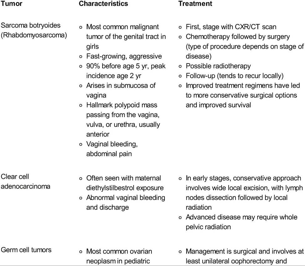
| Chlamydia trachomatis | ≤45 kg: erythromycin 50 mg/kg/d (divide in 4 doses/d) ? 14 d ≥45 kg, Sitz or tub baths twice a day for half an hour may help eliminate the vaginal discharge. • Nonirritating soaps and white cotton underpants should be recommended. • Nylon tights, tight blue jeans, prolonged wearing of wet bathing suits, and bubble baths should be discouraged. • Both the caregiver and child should be instructed on proper front to back wiping. • The child should be instructed to urinate with her knees apart to reduce urinary reflux into the vagina. • Persistent symptoms after therapy (>2 weeks) warrant reexamination. • In rare idiopathic persistent cases, vaginal irrigation with a 1% solution of povidone-iodine (Betadine) may help. • Alternate approaches to persistent cases include a 2-month course of antibiotics or 2 to 4 weeks of estrogen cream. • Recurrence often points to continued improper hygiene. Obese girls are at higher risk for recurrence. Prepubertal Vaginal Bleeding • Vaginal bleeding prior to menarche can result from a wide array of causes but must be taken seriously, as some conditions can be life-threatening. • Etiologies may include infection, anatomic abnormalities, genital tumors, hormonal abnormalities, trauma, or sexual abuse. Vulvovaginitis • Any cause of vulvovaginitis may result in vaginal bleeding. Evidence of infection, dermatoses, or retained foreign bodies should be targeted during evaluation. Urethral Prolapse • Increased abdominal pressure can cause the urethral mucosa to protrude through the meatus, forming an annular, hemorrhagic mass that bleeds easily. • Average age of onset is 5 years, and occurrence is more common in African Americans. • Medical treatment consists of a short-term course of estrogen cream. Topical antibiotics and sitz baths may also be beneficial. P.458 • Urinary retention or a large mass may require resection of the prolapsed tissue and insertion of an indwelling catheter. • Differential diagnosis includes urethral polyps, caruncles, cysts, and prolapsed ureteroceles. Genital Tumors • Genital tumors are uncommon in the prepubertal girl but need to be considered in a patient with a chronic genital ulcer, tissue protruding from the vagina, a malodorous or bloody discharge, or an atraumatic swelling of the external genitalia. • Causes are outlined in Table 34-2. Masses seen can be benign polyps or cancerous. Treatment is excision. • Sarcomas require a biopsy for diagnosis, excisional procedure, and chemotherapy. Abnormal Uterine Bleeding • See Chapter 40. Endometrial Shedding • Causes of endometrial shedding are outlined in Table 34-3 and often relate to a hormonal abnormality. • Precocious puberty is often associated with endometrial shedding in this population (see the section “Disorders of Puberty”). Trauma and Sexual Abuse • See the following text, also Chapter 33. Traumatic Injuries • The period of highest incidence is between ages 4 and 12 years, with 75% of all genital injuries occurring in young girls. Because of differences in anatomy between a child and an adult, a seemingly innocuous lesion can suggest serious injury. Common injuries include: Straddle Injuries • Most present as a swollen area of painful ecchymosis or hematoma over the labia; the mons, clitoris, and urethra can be involved. • If hematuria is present, consider a voiding Cystourethrogram to rule out bladder or urethral injury. • Periurethral injuries can result in swelling and urinary retention. Early placement of a urinary catheter is advised. • T reat with observation and cold compresses for the first 6 hours. If the hematoma remains the same size or becomes smaller, warm sitz baths are often all that are required. • Analgesics and prophylactic antibiotics can be used when a hematoma at the urethral orifice is causing pain and poor urination. Accidental Penetration • Most frequently seen between ages 2 and 4 years, often the result of falling on a sharp object (e.g., pen or pencil). • Presentation often includes hematuria, vaginal discharge, or bleeding. A puncture wound may be intraperitoneal with rectal pain or bleeding as the presenting complaint. • In an unstable patient with an injury above the hymen, laparoscopy or laparotomy should be performed. P.459 TABLE 34-2 Malignant Genital Tumors in Pediatric Gynecology
CXR, chest radiograph; CT, computed tomography; AFP, alpha-fetoprotein; hCG, human chorionic gonadotropin; CEA, carcinoembryonic antigen. P.460 TABLE 34-3 Causes of Endometrial Shedding in Children î Physiologic neonatal withdrawal bleed in the first 2 wk of life secondary to maternal estrogen withdrawal î Isolated premature menarche î Iatrogenic or factitious precocious puberty caused by medications that contain exogenous estrogens î Idiopathic precocious puberty î Functional ovarian cysts î Ovarian neoplasms î McCune-Albright syndrome î Central nervous system lesions î Hormone-producing neoplasms î Hypothyroidism • Workup involves examination with abdominal radiography, anoscopy, and sigmoidoscopy. Microscopic hematuria warrants careful urethral catheterization. Resistance to the passage of a catheter requires a voiding cystourethrogram. Catheterization should not be attempted with gross hematuria. Lacerations • Often secondary to forceful abduction of the legs, gymnastic exercise, water-skiing, bicycle accidents, or motor vehicle accidents. • Lacerations of the vaginal orifice frequently extend into the fornix. • Examination under anesthesia must be performed to determine the extent of the injury and rule out involvement of the rectovaginal septum or peritoneal cavity. Clitoral Strangulation or Ischemia • Difficult to diagnose; symptoms may include irritability and engorgement and cellulitis of the clitoris. • Often results when an entrapped hair from a caretaker accidentally becomes wrapped around the base of the organ. Treatment is removal of the stricture. Sexual Abuse • Suspect with unusual injury patterns or odd behavior as well as the following associated complaints: genital trauma, bleeding, chronic genital pain, sexually transmitted infections, anal inflammation, recurrent urinary tract infections, abdominal pain, enuresis/encopresis, or anorexia. • Behavioral changes include aggression, self-injury, conduct disorders, sleep disturbances, excessive phobias, depression, substance abuse, problems in school, or inappropriate knowledge of sexual behavior. • History: Obtain separately from the child if possible. Avoid leading questions. A doll may provide the young child with a way to express what has happened. A multidisciplinary approach involving the child's pediatrician and social worker may also be beneficial. • If abuse is suspected, the patient should be referred to an appropriate emergency department with individuals trained in collecting forensic evidence, preferably within 24 hours of the event. • Sexual play involves children of the same age without coercion and is a normal part of development. P.461 Labial Adhesions • In the low estrogen environment of childhood, the labia may fuse in response to any genital trauma, even diaper rash. • Adhesive vulvitis caused by chronic irritation is common between ages 2 and 6 years. • Asymptomatic labial adhesions do not require treatment and will resolve spontaneously with increasing estrogen levels in puberty. • If urinary retention or urinary tract infections occur, treatment is required and involves application of estrogen cream along the white line of the adhesion, with gentle traction twice daily for 2 to 6 weeks. • Recurrence is common after treatment. Acute urinary retention requires surgical excision. DISORDERS OF PUBERTY Puberty is a result of pulsatile gonadotropin-releasing hormone (GnRH) secretion and activation of the hypothalamic-pituitary-gonadal axis. The onset of puberty is generally between 8 and 13 years old in girls. Tanner stages are used to describe pubertal development. Delayed Puberty • Delay of puberty can be caused by anatomic abnormalities, chromosomal disorders, neoplastic growths, or nutritional deficiencies. • Commonly presents as a physical delay in maturation combined with amenorrhea. • Causes of delayed puberty can be classified based on the level of follicle-stimulating hormone (FSH) present, as outlined in Table 34-4. Hypergonadotropic Hypogonadism (High Follicle-Stimulating Hormone) • A sufficient amount of gonadotropins are present, but the ovaries are not responsive and therefore do not produce sex steroids. I I TABLE 34-4 An Overview of Causes of Delayed Puberty
|