Sexually Transmitted Diseases
Shireen Madani Sims
PELVIC INFLAMMATORY DISEASE
Any discussion of gynecologic infectious emergencies should begin with a review of pelvic inflammatory disease (PID). Approximately one million cases of PID per year result in 160,000 hospitalizations, and more than 100,000 surgical procedures annually (1).
Aside from the personal, physical, and emotional distress associated with PID, the financial cost to the individual and to society is staggering. In the United States, the total cost associated with PID and its sequelae in 2006 was estimated at $4.2 billion (1).It is important for the clinician to recognize certain risk factors that may be associated with an increased risk of PID. A major risk factor is age. Young women are at greater risk of acquiring PID as a consequence of the greater prevalence of sexually transmitted diseases, a lower prevalence of protective chlamydial antibodies, larger zones of cervical ectopy, and a greater penetrability of cervical mucus (2). Sexually active adolescents are three times more likely to be given the diagnosis of PID than women who are 25 to 29 years old (1). Multiple sexual partners, a high frequency of sexual intercourse, and a high rate of new partner acquisition within the previous 30 days all appear to increase the acquisition risk of PID (3). The perception of the role of an intrauterine device (IUD) and the risk of PID is undergoing review. The primary risk centers around the time of insertion (4). It is presumed to be secondary to the introduction of vaginal and cervical pathogens into the endometrium during IUD insertion. However, even in areas with high prevalence of STDs, the use of an IUD appears to only minimally increase the risk of PID (4,5). Other factors that appear to be associated with an increased risk of PID include douching, smoking, and proximity to menses. It has been observed that symptoms develop significantly more often in women with chlamydial or gonococcal salpingitis within 7 days of menses than at other times during the cycle (6).
Although Neisseria gonorrhoeae and Chlamydia trachomatis are considered to be the primary pathogens in PID, other organisms are also isolated from tubal and peritoneal fluids. These organisms include aerobes (Streptococcus species, Escherichia coli, and Haemophilus influenzae) and anaerobes (Bacteroides bivius, Bacteroides fragilis, Peptostreptococcus, and Peptococcus) (7). Other organisms that have been isolated but whose significance is still indeterminate include Mycoplasma and Actinomycosis. Finally, tuberculosis is a remote consideration in patients at risk (particularly in underdeveloped countries and immunosuppressed patients). Although PID may be caused by a single agent, infection is frequently polymicrobial. The polymicrobial nature of PID may result when more than one primary pathogen infects the oviductal epithelium or it may result from damage created by a single primary pathogen, resulting in altered host immune defense mechanisms and secondary infections by other bacteria. These secondary invading organisms may be normal inhabitants of the upper vagina that become pathogenic upon contact with damaged tubal epithelium.
PID has always posed a diagnostic dilemma for the examining physician. Mild disease may be misdiagnosed as cystitis or gastroenteritis. Severe disease may be misdiagnosed as ovarian torsion, diverticulitis, or, most commonly, appendicitis. Frequently, this diagnostic dilemma results from the lack of sensitivity and specificity of the patient’s complaints, physical findings, and laboratory evaluation. Therefore, the index of suspicion for the clinical diagnosis of PID should be high. The CDC has recommended a minimal set of criteria for empiric treatment in order to reduce missed or delayed diagnosis (8,9). It may be appropriate to overtreat as undertreatment may cause significant damage to the oviducts, resulting in chronic pelvic pain, infertility, or a future ectopic pregnancy. When no other cause can be identified, empiric treatment should be started in women with pelvic pain and at least one of the following: (a) cervical motion tenderness or uterine/adnexal tenderness; (b) oral temperature >101°F (>38.3 °C); (c) peripheral leukocytosis or left shift; (d) abnormal cervical or vaginal mucopurulent discharge; (e) presence of white blood cells (WBCs) on saline microscopy of vaginal secretions; (f) elevated erythrocyte sedimentation rate; and (g) elevated C-reactive protein.
Patients with pelvic pain and any of the following are considered confirmed cases: (a) endometrial biopsy with histopathologic evidence of endometritis; (b) laparoscopy findings consistent with PID; (c) laboratory evidence of cervical infection with N. gonorrhoeae or C. trachomatis; and (d) imaging studies demonstrating thickened, fluid-filled oviducts with or without free fluid or tubo-ovarian complex.The use of ultrasonography and computed tomography (CT) scanning may be useful in examining the patient with severe rebound tenderness in whom an adequate pelvic examination is impossible to perform. Ultrasonography may demonstrate echogenic fluid in the pelvis consistent with pus (Fig. 19.1). CT scanning may demonstrate abscess formation with fluid and air collection (Fig. 19.2).
When significant uncertainty exists concerning the diagnosis, the “gold standard” has been the performance of a diagnostic laparoscopy. One major study of patients with signs and symptoms of PID undergoing a laparoscopic examination demonstrated that in 65% of the patients, the correct preoperative diagnosis had been made, 23% had normal pelvic anatomy, and 12% had other pathology (appendicitis or endometriosis) (10).
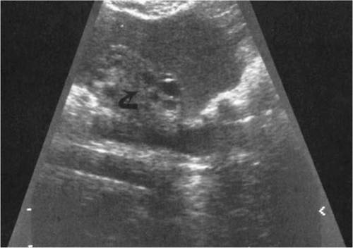
FIGURE 19.1 Transvaginal probe demonstrating ovary with follicles [arrow] surrounded by echogenic fluid (pus).
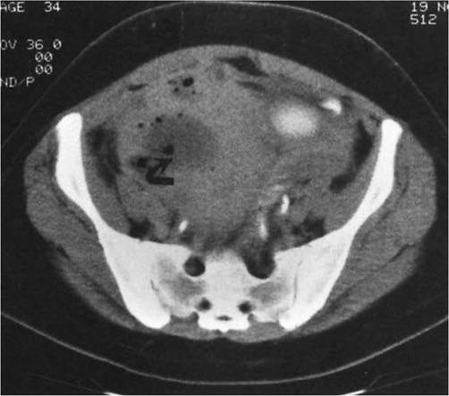
FIGURE 19.2 CT scan demonstrating tubo-ovarian abscess with fluid collection and gas (arrow).
The decision to admit the patient to the hospital for further evaluation or treatment should be based on certain well-established criteria. These include the following: (a) significant peritoneal signs or rebound tenderness, (b) presence of an IUD, (c) pregnancy, (d) an adnexal mass consistent with a tubo-ovarian abscess (on pelvic examination or diagnostic imaging), (e) gastrointestinal symptoms precluding appropriate outpatient therapy or suggestive of bowel pathology, (f) failed outpatient therapy, (g) nulliparity, and (h) an uncertain diagnosis.
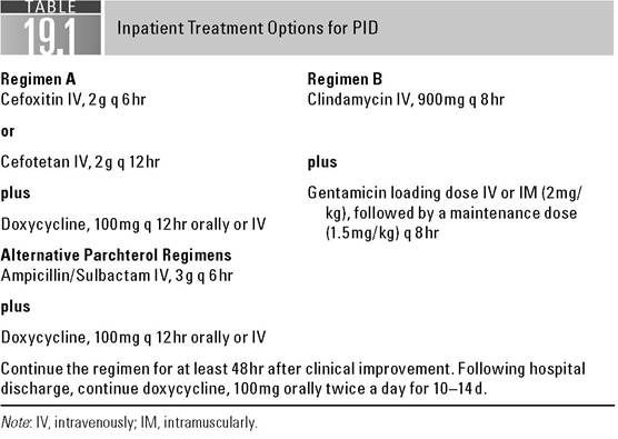
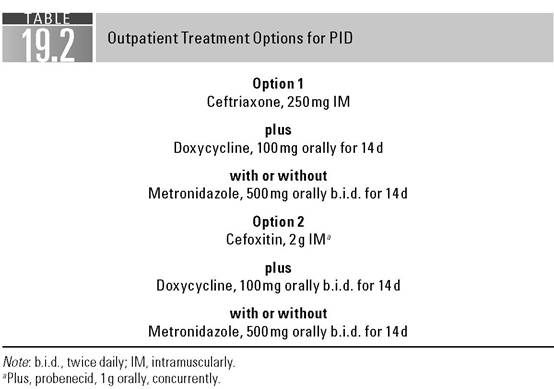
Patients with an uncertain diagnosis require further evaluation and institution of therapy. The decision to perform laparoscopy to delineate the disease process may be based on the patient’s severity of symptoms on admission or on her response to antibiotic therapy. If the decision is to initiate treatment with antibiotics and no resolution of symptoms occurs in 24 to 48 hours, or if symptoms increase in severity during this time frame, a laparoscopy should be considered. Treatment regimens for PID are outlined in Tables 19.1 and 19.2. Appropriate follow-up ensures compliance and resolution of symptoms in patients undergoing the outpatient regimen.Ruptured tubo-ovarian abscess or leaking tubo-ovarian abscess may present as a serious threat to life. Mortality rate associated with a ruptured tubo-ovarian abscess ranges from 10% to 15%. Appropriate diagnosis and therapy are necessary. When a tubo-ovarian abscess ruptures, endotoxin is released into the systemic circulation. The lipid A portion of the lipopolysaccharide results in the release of numerous factors or mediators: ^-endorphins, bradykinin, activators of the complement and coagulation cascades, plasminogen, and histamine. Decreased systemic resistance, a low pulmonary artery occlusive pressure, and increased cardiac output are noted in the initial stage of shock. As shock progresses, systemic vascular
resistance increases and cardiac output decreases. Microvascular hypoperfusion resulting from microembolization from fibrin degradation products and precapillary sphincteric dilatation resulting from hypoxia result in arterial venous shunting and loss of effective intravascular volume. Myocardial depression results in a diminution of effective cardiac output. Inadequate perfusion of vital organs results in renal dysfunction and accentuated acidosis.
It is imperative that the managing physician recognize the presence of septic shock and respond appropriately. 'The hypotensive, tachycardic patient with abdominal findings suggestive of diffuse peritonitis, secondary to a ruptured tubo-ovarian abscess, should be managed aggressively with fluid resuscitation. Large volumes of crystalloid should be administered intravenously through two large-bore catheters. If there is a failure to correct hypotension with fluid administration, intravenous sympathomimetic amines should be initiated. Dopamine is the primary agent to be considered. Administered at a dose of 1 to 3 μg∕kg per minute, dopamine has minor inotropic and chronotropic effects on the heart, with concomitant dilatation of mesenteric, cerebral, coronary, and renal arteries (11,12). At dosages between 4 and 10 μg∕kg per minute, there is a further increase in cardiac output and an increased heart rate. The decision to initiate invasive monitoring for fluid resuscitation should be based on the patient’s response to crystalloid administration and urine output. If blood pressure fails to respond to intravascular volume repletion and sympathomimetic amines and if urine output is not appropriate (>30 mL per hour), then pulmonary capillary wedge pressure monitoring should be considered to prevent the development of adult respiratory distress syndrome. Broad-spectrum antibiotic therapy should be initiated promptly to cover the polymicrobial gamut of pelvic pathogens. Preparation should be made for emergency admission to the hospital and laparotomy to remove the ruptured abscess and irrigate the peritoneal cavity. Blood should be sent for type and cross-match, coagulation panel, electrolytes, and blood gases. It is imperative that the physician managing the patient in septic shock from a ruptured tubo-ovarian abscess understand that this is a surgical disease, and pharmacologic intervention is supportive and preparatory to laparotomy.GONORRHEA
Neisseria gonorrhea requires emergency care in two situations: (a) when it is associated with acute PID and (b) when it is the cause of a disseminated gonococcal infection.
Only 1% to 2% of patients with gonorrhea develop disseminated gonococcal infections. Women appear to be more commonly affected by this presentation than men. This may be explained by the relative lack of symptoms in women harboring N. gonorrhea in the lower reproductive tract, specifically the cervix. With a breakdown in host immune defense, gonococcemia may occur. With the invasion of the bloodstream, symptoms include fever, chills, and arthralgias. Gonococcal dermatitis-arthritis syndrome develops within 2 to 3 weeks of the primary genital infection. Cutaneous manifestations usually consist of fewer than 25 lesions, usually on the distal extremities and in various stages of development. Lesions begin as pinpoint erythematous macules, which progress to papules, vesicular pustules, or hemorrhagic bullae. Advanced lesions contain a necrotic-appearing center surrounded by an erythematous halo. Rarely are cultures of cutaneous lesions positive for N. gonorrhea; however, immunofluorescent tissue stains may be of assistance in demonstrating organisms. Blood, urethral, cervical, pharyngeal, and rectal cultures may be of assistance in defining the etiology of the rash. A high index of suspicion is imperative (13).
With dissemination, the patient may have an acutely inflamed septic joint. Disseminated gonococcal disease is the most common cause of septic arthritis in patients younger than 30 years of age. The arthritis may be monoarticular or oligoarticular. Joints most commonly affected are the knees, elbows, ankles, wrists, and small joints of the hands and feet (14). The knee is the most commonly involved joint from which the gonococcus is recovered, but this may reflect the relative ease with which this joint is aspirated. The fluid withdrawn from the involved joint usually contains polymorphonuclear neutrophils, but a Gram stain for Gram-negative intracellular diplococci is positive only 10% to 30% of the time. Cultures likewise are positive in approximately 20% to 30% of cases. Once again, it is imperative that appropriate cultures be obtained from blood, urethral, pharyngeal, rectal, and cervical sites.
With the dissemination of the gonococcus to the heart, endocarditis may develop. Patients may have fever, chills, arthralgias, malaise, fatigue, dyspnea, and chest pain. Most patients have a murmur, and evidence of embolization may be present (conjunctival petechia, Osler’s nodes, and splinter hemorrhages). Occasionally, splenomegaly and arthritis may be noted. Depending on the degree of cardiac compromise, congestive heart failure may be manifested by rales, ascites, edema, or a gallop rhythm on auscultation of the heart. The chest x-ray film may demonstrate cardiomegaly as a manifestation of congestive heart failure. The electrocardiogram may demonstrate left ventricular hypertrophy, bundle branch block, or intraventricular conduction delay. Usually, blood cultures are positive for N. gonorrhea. Echocardiography is useful in determining whether vegetations exist on the heart valves. The most common involvement is of the aortic and mitral valves. Without treatment, the endocarditis is almost always fatal.
Rarely, N. gonorrhea may disseminate to the meninges and cause manifestations of meningitis. Fever and nuchal rigidity in a patient complaining of headache, general malaise, and arthralgias should prompt a lumbar puncture to evaluate for organisms. As with synovial fluid, Gram stain and cultures may be negative, and therefore it is imperative to perform appropriate blood, cervical, urethral, rectal, and pharyngeal cultures for Neisseria.
Hospitalization is recommended for patients with disseminated gonococcal infection. Endocarditis and meningitis should be ruled out. Ongoing CDC investigation demonstrates that fluoroquinolone-resistant gonorrhea is now widespread in the United States. Therefore, this class of antibiotic is no longer recommended for treatment. When bacteremia and arthritis are present, the recommended therapy is ceftriaxone, 1 g intravenously (IV) daily for 7 to 10 days. Meningitis should be treated with ceftriaxone, 1 to 2 g IV every 12 hours for at least 10 days. Endocarditis should be treated with ceftriaxone, 1 to 2g IV every 12 hours for at least 4 weeks. Depending on the severity of the valvular involvement with vegetations, cardiac surgery with valvular replacement may be necessary. Consultation with the appropriate specialist should be obtained early (15,16).
ACQUIRED IMMUNODEFICIENCY SYNDROME
Even though the decade of the 1980s began with human immunodeficiency virus (HIV) as a male-specific entity, this virus is clearly not gender specific. The estimated number of new infections in the United States in 2006 was 56,300. Women account for 25% of patients living with HIV, with a prevalence of 278,400 in 2006 (17). The gynecologist cannot avoid the potential for exposure to patients infected with HIV, whether or not the patient’s infectivity status is known.
HIV is an RNA retrovirus, which uses reverse transcriptase to transcribe DNA from RNA. It targets the CD4 molecule on the surface of the T4 lymphocyte. After being incorporated into the T4 lymphocyte and using reverse transcriptase to transcribe DNA, the virus is integrated into the host genome and the production of viral RNA begins. T4 cells are comprised of inducer and helper cells. Inducer cells stimulate the maturation of T lymphocytes from precursor cells, and T4 helper cells help cytotoxic T cells destroy foreign cells. T4 cells constitute approximately 60% to 80% of the circulating T-cell population. As a result of the infection and depletion of T4 lymphocytes by the invading HIV the B cells cannot produce antibodies to HIV or other microorganisms. The cytotoxic response is depressed. There is a decreased secretion of interleukin-2, and T4 cells are incapable of antigen recognition. Not only does HIV attack the T helper cells but also the virus may attack macrophages and other target cells, resulting in direct infection of bowel, nervous tissue, heart, and lung (18). It is well recognized that the infected host may transmit HIV to susceptible individuals through blood or body fluids, including vaginal secretions, semen, peritoneal fluid, and amniotic fluid (19). Once individuals are infected with HIV, seroconversion may not occur for a mean of 18 months, with a range of 3 to 42 months (20). The mean latency period from seroconversion to AIDS is usually 10 to 11 years. Once AIDS develops, death is inevitable. As the disease progresses in the female patient, a number of opportunistic infections may result in infections that prompt the need for emergent therapy.
Because AIDS is a sexually transmitted disease, the AIDS patient is prone to acquisition of other sexually transmitted diseases, and it is important to keep this in mind when evaluating them. For example, an AIDS patient with signs and symptoms of PID may not demonstrate leukocytosis. For this reason, strong consideration should be given to admitting the AIDS patient with signs and symptoms of PID for intravenous antibiotic therapy. Currently, there appears to be no basis to provide treatment regimens other than those currently recommended by the Centers for Disease Control for acute PID (9).
The observant physician should be alert to clinical manifestations of AIDS in any patient seen in an emergency setting. Such manifestations include enlarged lymph nodes, night sweats, fevers, oral candidiasis, chronic cough, paresthesias, nausea, vomiting, diarrhea, weight loss, and skin ulcerations.
The primary pulmonary emergency in the patient with AIDS is Pneumocystis jiroveci (formerly Pneumocystis carinii) pneumonia. This most common of the opportunistic infections in patients with AIDS poses the greatest risk to those who have CD4 T-lymphocyte counts of whether or not serum status is known.
SYPHILIS
Treponema pallidum was responsible for epidemic outbreaks of syphilis in the 15th and 16th centuries in Europe. Widespread prevalence of the disease was observed until the availability of penicillin after World War II. In 1943, 575,000 cases were reported in the United States (26). In 2000, the rate was the lowest since reporting began in 1941. However, a disturbing trend emerged, with the rate of syphilis increasing steadily between 2000 and 2007, with 40,920 cases reported in 2007 (27).
T. pallidum is a delicate, spiral-shaped organism, which is spread by sexual contact. After inoculation, the average incubation period is 3 weeks, with a range of 9 to 90 days. The initial lesion of syphilis, known as the primary lesion or chancre, is usually found in the genital region, although extragenital chancre may occur on the lip or finger (Fig. 19.5). The chancre is a firm, well-circumscribed painless ulceration with a clean base. If superinfection is present, the ulceration may be painful. Regional painless lymphadenopathy is also observed. The chancre may persist for 3 to 8 weeks and then heal spontaneously; however, the lymphadenopathy may persist for longer durations. Without therapy, within weeks to months, the stage of secondary syphilis is entered. Approximately 50% of patients demonstrate signs and symptoms during this stage, which may consist of low-grade fever, meningismus, arthralgia, nocturnal bone pain, coryza, and malaise. The painless chancre of primary syphilis may go unobserved, especially if it is located on the cervix, and therefore women often do not seek evaluation until they have entered this stage. Skin lesions in this stage of the disease may be annular, maculopapular, papular, or macular. The most common skin lesion is the maculopapular syphilid, which occurs in approximately 70% of cases and consists of distinctive brownish or coppery macules and papules on the palms and soles (Fig. 19.6). Another characteristic lesion of secondary syphilis is condyloma latum, which is often confused with condyloma acuminata
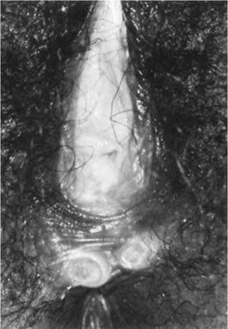
FIGURE 19.5 Primary chancres.
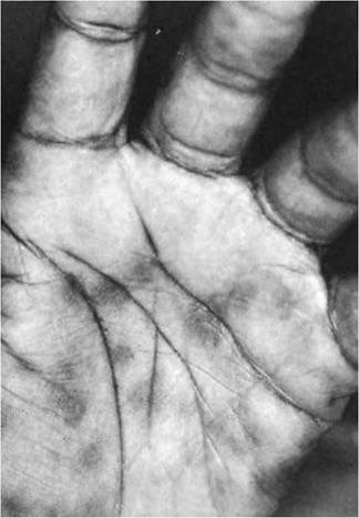
FIGURE 19.6 Maculopapular secondary syphilis.
induced by human papillomavirus. This lesion is broad based, moist, and grayish white in coloration. It is highly contagious and may also be found in other warm moist regions of the body, such as the inner thighs, the axilla, and underneath the breasts. Dark-field examination, serologic testing, or biopsy may confirm the diagnosis.
If the Treponema organisms induce osteomyelitis that spreads to the periosteum, patients may complain of bone pain, which classically is nocturnal and exaggerated by heat, frequently being relieved by movement of the involved bone (28). The skull, clavicle, tibia, and radius are the most commonly involved bones in patients with periostitis as a manifestation of secondary syphilis. This bone pain may precede or follow the mucocutaneous manifestations of secondary syphilis and may be the only manifestation of secondary syphilis.
The liver may be involved in secondary syphilis, and patients may complain of abdominal pain and upper quadrant discomfort. Approximately 50% of patients with secondary syphilis may have liver function abnormalities, and 1% to 12% may demonstrate jaundice. Most cases of hepatic dysfunction, during the course of early syphilis, are a result of inflammation of the liver by the spirochetes. It has been suggested that rectal inoculation with T pallidum results in direct portal venous inoculation and a higher incidence of hepatic involvement (29).
Ocular involvement in secondary syphilis may result in a patient seeking care for visual disturbances. The pathogenesis of syphilitic episcleritis and scleri- tis is related to lymphocytic infiltration and subsequent vasculitis. Patients may have a painful, reddened eye. Iritis presenting as pain, photophobia, and dimness of vision may be noted, and adhesions of the iris to the anterior lens may produce a fixed pupil. This is not to be confused with the Argyll Robertson pupil, which is also seen as a component of symptomatic neurosyphilis and presents as a small irregular pupil that reacts to accommodation but not to light (30).
The central nervous system is commonly involved in early syphilis, affecting at least 50% of patients (31). Although central nervous system involvement is frequent, symptomatic neurosyphilis develops in fewer than 10% of untreated patients. Patients with symptomatic neurosyphilis may have cranial nerve palsies, paresthesias, weakness, sensory loss in extremities and trunk, progressive mental deterioration, convulsions, ataxia, areflexia, bladder and eye disturbances, optic atrophy, coma, and death. These manifestations of syphilis are typically seen in late syphilis, particularly in patients not treated for primary or secondary syphilis. Patients with these symptoms should be evaluated for the possibility of untreated or suboptimally treated neurosyphilis.
The evaluation of a patient with the aforementioned symptoms suggestive of syphilis is well established. The lesions should be examined for spirochetes. Obvious care should be exercised, because mucocutaneous lesions are infectious. Serology is likewise helpful, but because primary syphilis manifests as a chancre, initial serology may be positive in 70% of patients and negative serologic examination in the absence of the ability to perform a dark-field examination does not exclude the diagnosis of primary syphilis. Antibodies against cardiolipin, known as reaginic or nontreponema antibodies (VDRL [Venereal Disease Research Laboratory] and RPR [Rapid Plasma Reagin]), are quantitative measurements and serum titers usually reflect disease activity. The specific treponemal antibody test, the fluorescent treponemal antibody absorbed test (FTA-ABS), is very sensitive and specific and usually remains reactive indefinitely without regard to treatment status. This test is useful in ruling out false-positive, nontreponemal tests, which may be caused by pregnancy, recent immunizations, sarcoidosis, hepatitis, and autoimmune diseases such as systemic lupus erythematosus. For the patient with a lesion of primary syphilis, the initial screen should include a dark-field examination of the chancre, qualitative and quantitative nontrepone- mal assays (VDRL or RPR), and a specific treponemal assay (the FTA-ABS). The differential diagnosis of such a lesion includes HSV, chancroid, and HIV. Appropriate cultures for Haemophilus ducreyi and HSV should be obtained if suspected, and HIV testing should be considered. Syphilis is the great masquerader, and even though it may be highly likely that secondary syphilis will be suspected in patients with maculopapular diffuse eruptions involving the palms and soles, the diagnosis must also be entertained in patients with right upper quadrant pain, bone pain, ocular symptoms, and neurologic symptoms. The most consistently observed laboratory abnormality in syphilitic hepatitis is a marked elevation in alkaline phosphatase. The differential diagnosis should include viral hepatitis, cholecystitis, and Fitz Hugh-Curtis syndrome. In addition to the liver function tests, studies should be obtained for hepatitis B and treponemal and nontreponemal serologies. Patients with bone pain may demonstrate tenderness on physical examination while the involved bone is palpated. In the absence of obvious mucocutaneous lesions of secondary syphilis for dark-field studies, the VDRL and FTA-ABS should be obtained. The erythrocyte sedimentation rate may be elevated. Radiographs may show no radiologic changes. Bone scanning may show increased uptake in the involved bones suggestive of inflammation. Patients with syphilitic scleritis typically have positive nontreponemal and treponemal serologies. Other causes for the inflammation should be evaluated with assays for rheumatoid factor and antinuclear antibody.
Lumbar puncture is recommended to rule out neurosyphilis in all patients with syphilis of more than 1 year’s duration or in patients with clinical symptoms of meningitis or focal neurologic findings. The issue of whether all patients should have a lumbar puncture remains unanswered; however, it has been observed that patients with concurrent HIV infection and positive central nervous system serology have a higher failure rate on standard treatment with the usually recommended dose of 2.4 million units of benzathine penicillin as a single-dose therapy for primary syphilis. Until the optimum therapy for HIV- testing for HIV in all patients with syphilis. Lumbar puncture should be strongly considered in patients who are HIV positive and seropositive for syphilis before the initiation of therapy (32).
Therapy for early syphilis (primary syphilis, secondary syphilis, and latent syphilis of the patient unable to tolerate oral medication, and the patient with disseminated mucocutaneous HSV need to be hospitalized. Acyclovir should be administered intravenously at 5 mg/kg every 8 hours for 5 to 7 days (38). In AIDS patients with severe herpes genitalis, strong consideration should be given to hospitalization for intravenous therapy (39).
Certainly, patients with nonprimary first-episode herpes or patients with recurrent herpes may have significant discomfort from their disease; however, attacks are less likely to be associated with severe systemic symptoms or urinary retention. Patients with nonprimary first-episode herpes have circulating antibodies from a prior herpes infection that usually limit the manifestations of the disease. The same can be said for patients who have episodes of recurrent herpes virus infections. The diagnosis is suspected after observing the genital lesion, which is typically ulcerative when first seen by the physician. The patient may have noted the erythematous papule developing into a painful vesicle, usually within the first day of a recurrent infection. With progression, the vesicle ruptures and ulceration forms. Viruses are recoverable from the lesion between the first and the fourth days of recurrence. Recovery rates are lower with each stage of the disease than in primary infections. Treatment of recurrent genital herpes with acyclovir, 200mg orally five times a day for 5 days, has been shown to reduce viral shedding by 1 day and shorten the duration of the episode by 1 to 2 days (40). It is imperative that therapy for recurrence be initiated early in the course of the disease or the ameliorative effect is lost.
CHANCROID
Chancroid is a relatively infrequent disease in the United States, accounting for 33 cases in 2006 (41). The disease is caused by H. ducreyi, a Gram-negative facultative anaerobic bacillus. After an incubation period of 4 to 7 days, a chancre appears, which rapidly erodes to form an ulcer approximately 48 hours after its initial appearance. The ulcer is extremely tender, with irregular undermined edges. The base of the ulcer is necrotic, whereas the syphilitic chancre is clean, without evidence of necrosis. Multiple lesions develop in 33% of patients, and contiguous lesions may coalesce into giant ulcers. Lesions in women are most common on the fourchette, the labia, the vestibule, and the clitoris. Without treatment, extensive ulceration and massive edema of the vulva and perineum may be observed. Inguinal lymphadenopathy develops in half of patients, and these may develop into a bubo with rupture and adjacent ulceration.
The diagnosis of chancroid is suspected when the characteristic lesion is noted. Gram stain from the base of the ulcer shows Gram-negative coccobacil- lary forms in chains. Special culture media (gonococcal agar base and Mueller Hinton agar base) can be used to culture for the organism. Strong consideration should be given to HIV testing, because there is an increased rate of chancroid infection among patients with HIV.
The recommended regimen for the treatment of chancroid is either azithromycin, 1g orally once, or ceftriaxone, 250mg intramuscularly once. Other regimens include ciprofloxacin, 500 mg orally twice a day for 3 days, or erythromycin, 500 mg orally four times a day for 7 days (42). With successful treatment, ulcers will improve within 3 days symptomatically and within 7 days objectively. If no clinical improvement is observed by 7 days, the clinician should determine whether the prescribed medication was taken and rule out coinfection with another sexually transmitted disease such as syphilis or herpes, in addition to ruling out infection with HIV. A final consideration would be antibiotic resistance, and appropriate susceptibilities should be determined.
References
1. www2a.cdc∕gov∕std. Last accessed 6/09.
2. Cates W, Rolfs RT, Aral SO. Sexually transmitted diseases, pelvic inflammatory disease, and infertility: an epidemiologic update. Epidemiol Rev. 1990;12:199-220.
3. Washington EA, Aral SO, Wolner-Hanssen P, et al. Assessing risk for pelvic inflammatory disease and its sequelae. JAMA. 1991;266:2581-2586.
4. Meirik O. Intrauterine devices—upper and lower genital tract infections. Contraception. 2007;75:S41-S47.
5. Shelton JD. Risk of clinical pelvic inflammatory disease attributable to an intrauterine device. Lancet. 2001;357:443.
6. Sweet EL, Blankfort-Doyle M, Robbie MO, et al. The occurrence of chlamydial and gonococcal salpingitis during the menstrual cycle. JAMA. 1986;255:2062-2064.
7. Rice PA, Schachter J. Pathogenesis of pelvic inflammatory disease: diagnostic and prognostic value of routine laparoscopy. Am J Obstet Gynecol. 1969;105(7): 1088-1098.
8. Kahn JG, Walker CK, Washington E, et al. Diagnosing pelvic inflammatory disease. JAMA. 1991;266:2594-2604.
9. Workowski KA, Berman SM. Sexually transmitted diseases treatment guidelines, 2006. MMWR Recomm Rep. 2006;55(RR-11):1-94.
10. Jacobson L, Swastrom L. Objectivized diagnosis of acute pelvic inflammatory disease: diagnosis and prognostic value of routine laparoscopy. Am J Obstet Gynecol. 1969;105:1088-1098.
11. Drugs for sexually transmitted infections. Med Lett. 1999;41(1062):85-90.
12. Septic shock: Where are we now? Emerg Med Rep. 1991;33:856.
13. Buntin DM, Rosen T, Lesher JL, et al. Sexually transmitted diseases: bacterial infections. JAm Acad Dermatol. 1991;25(2) 287-299.
14. Dallabetta G, Hook E. Gonococcal infections. Infect Dis Clin North Am. 1987;1:25-54.
15. Wall TC, Peyton RB, Corey GR, et al. Gonococcal endocarditis: a new look at an old disease. Medicine. 1989;68:375-380.
16. Update to CDC's sexually transmitted diseases treatment guidelines, 2006: fluoroquinolones no longer recommended for treatment of gonococcal infections. Morbid Mortal Wkly Rep. 2007;56(14):332-336.
17. Hall HL, Song R, Rhodes P, et al. Estimation of HIV incidence in the United States. JAMA. 2008;300(5):520-529.
18. Levy JA. Changing concepts in HIV infection: challenges for the 1990's. AIDS. 1990;4:1051.
19. US Department of Health and Human Services, Public Health Services, Centers for Disease Control. Acquired immunodeficiency syndrome United States, 1989. Morbid Mortal Wkly Rep. 1990;39(5):81.
20. Wolinsky SM, Rinaldo CR, Kowk S, et al. Human immunodeficiency virus type 1 (HIV-1) infection in a median of 18 months before a diagnostic Western blot: evidence from a cohort of homosexual men. Ann Intern Med. 1989;111:961.
21. Drugs for AIDS and associated infections. Med Lett. 1995;37:(959):87-94.
22. Briel M, Bucher HC, Furrer H. Adjunctive corticosteroids for Pneumocystis jiroveci pneumonia in patients with HIV-infection. Cochrane Database Syst Rev. 2006 Jul;3:CD006150.
23. Dwyer B. HIV in the 2nd decade: clinical manifestations and emergency management. Emerg Med Rep. 1991;12:22.
24. Covino JM, McCormach WM. Vulvar ulcer of unknown etiology in a human immunodeficiency virus-infected woman: response to treatment with zidovudine. Am J Obstet Gynecol. 1990;163:1.
25. Henderson DK, Fahey BJ, Willy M, et al. Risk for occupational transmission of human immunodeficiency virus type I (HIV-1) associated with clinical exposures. Ann Intern Med. 1990;113:740.
26. CDC. Sexualty dransmittedDisease Statistics—1987. Atlanta, GA: Centers for Disease Control; 1988:136;988.
27. CDC. Sexually Transmitted Disease Surveillance-2007. Atlanta, GA: Centers for Disease Control.
28. Meier JL, Mollet E. Acute periostitis in early acquired syphilis simulating shin splints in a jogger. Am J Sports Med. 1986;14(4):327-328.
29. Sclossberg D. Syphilitic hepatitis: a case report and review of the literature. Am J Gastroenterol. 1987;82(6):552-553.
30. Wilhelmus K, Yokoyama C. Syphilitic episcleritis and scleritis. Am J Ophthalmol. 1987;104:595-597.
31. Zenker RN, Rofs RT. Treatment of syphilis, 1989. Rev Infect Dis. 1990;12:5590-5609.
32. Lukehart SA, Hook EW, Baker-Zander SA, et al. Invasion of the central nervous system by Treponema pallidum: implications for diagnosis and treatment. Ann Intern Med. 1988;109:855-862.
33. Centers for Disease Control and Prevention. 2006 Guidelines for Treatment of Sexually Transmitted Diseases. Morbid Mortal Wkly Rep. 2006; 55:22.
34. Rosen T, Rubin H, Ellner K, et al. Vesicular Jarisch-Herxheimer reaction. Arch Dermatol. 1989;125:77-81.
35. Landy HJ, Grossman JH. Herpes simplex virus: sexually transmitted diseases. Obstet Gynecol Clin North Am. 1989;16(3):495-515.
36. Gutmann D, Beard BA, Collinge ML, et al. Nonfatal disseminated mucocutaneous herpes simplex type 2 infection in a healthy woman. Obstet Gynecol. 1988;72:506- 507.
37. Webb D, Fife K. Genital herpes simplex virus infections: sexually transmitted diseases. Infect Dis Clin North Am. 1987;(1):1:97-122.
38. Drugs for sexually transmitted infections. Med Lett. 1999;41(1062):85-90.
39. Kroon S. Genital herpes—When and how to treat. Semin Dermatol. 1990;9(2): 133-140.
40. Stone K, Whittington W. Treatment of genital herpes. Rev Infect Dis. 1990;12(6): 610-619.
41. Centers for Disease Control and PRevention, Division of SExually Transmitted Diseases. Sexually Transmitted Diseases Surveillance, Other Sexually Transmitted Diseases, 2006 National Report. Available at: http://www.cdc.gov/std/stats/other. htm. Accessed May 7, 2008.
42. Centers for Disease Control and Prevention. 2006 Guidelines for Treatment of Sexually Transmitted Diseases. Morbid Mortal Wkly Rep. 2006;55:15.