Cerebellum and Hindbrain
The most common hindbrain abnormality in neural tube defects is Chiari type II malformation, seen in 80% to 90% of individuals with myelomeningocele (23,24,27,51).
This malformation results in caudal displacement or herniation of the medulla, lower pons, elongated fourth ventricle, and cerebellar vermis into the cervical spinal cord (Fig.
9.4). This often interferes with cerebrospinal fluid outflow and is, therefore, almost always associated with hydrocephalus. Caudal displacement of the medulla may occur and result in traction neuropathies of the lower cranial nerves. Signs of bulbar compromise arise from compression of the herniated hindbrain.A broad spectrum of clinical symptoms is seen in individuals with this malformation. However, only 20% will develop clinical signs of brainstem dysfunction, with most occurring in the neonatal period (52,53). Symptoms may be evident at birth or present within the first two to three months.
The most severe symptom is respiratory compromise, which may be both central and peripheral in
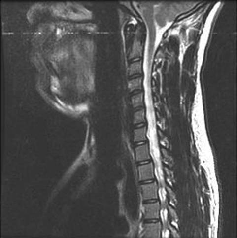
Figure 9.4 T2-weighted magnetic resonance image of the cervical spine. The posterior fossa is crowded. There is cerebellar tonsillar herniation, with the cerebellar tonsils lying 9 millimeters below the foramen magnum. This is the expected finding for a Chiari II malformation.
etiology. Individuals may experience stridor, laryngeal nerve palsy with vocal cord paralysis, upper airway obstruction, periodic breathing, central or obstructive sleep apnea, or aspiration. Dysphagia and extraocular motion abnormalities may also be seen related to other cranial neuropathies. Dysphagia may be severe enough that gastrostomy tube placement is required. Airway compromise may necessitate tracheostomy.
In the presence of brainstem compromise, hemiparesis or tetraparesis may be seen (this is more common in older children or adults than infants). Impairment of fine motor hand function is well documented and is seen in more than half of individuals with thoracic- level lesions and approximately one-fourth of individuals with lumbosacral lesions.
Control of ocular motility is related to cerebellar function (saccadic eye movements, visual fixation, and pursuit). There is a high rate of visual problems in individuals with spina bifida. Fewer than one-third have completely normal visual function (54,55).
Despite successful initial treatment with surgical decompression, problems may recur. Typically, vocal cord paresis in the first two months of life is a sign of irreversible damage, and surgical decompression is unlikely to result in clinical improvement (56).