Congenital Muscular Dystrophy
The term congenital muscular dystrophy (CMD) has been widely used for a group of infants presenting with hypotonia, muscle weakness at birth or within the first few months of life, congenital contractures, and immunohistochemical finding of dystrophic changes on muscle biopsy (muscle fiber necrosis and regeneration, increased endomysial connective tissue, and replacement of muscle with fat tissue).
The early contractures may include equinovarus deformities, knee flexion contractures, hip flexion contractures, and tightness of the wrist flexors and long finger flexors. The contractures can become more severe over time with prolonged static positioning and lack of adequate passive range of motion and splinting/posi- tioning. Classical CMDs are clinically confined to the musculoskeletal system, but other CMDs are characterized by significant cerebral neuronal migration defects and eye abnormalities. Classical CMDs are further subdivided according to the presence or absence of merosin (laminin-2) (64). An additional subgroup with collagen VI abnormalities has been identified and referred to as Ullrich’s congenital muscular dystrophy.Merosin-Deficient CMD
This condition (CMD 1A) has been linked to chromosome 6q22 and accounts for around half of classical CMD (64). These children show a consistently severe phenotype with multiple contractures and joint deformities (arthrogryposis) at birth. Weakness correlates with level of residual merosin (laminin α2) protein. If there is absent laminin α2 protein, weakness is severe, symmetric, proximal greater than distal, and involves the facial muscles. Contractures are present at multiple joints. CK is mildly to moderately elevated. Infants may present with respiratory failure, but if adequately supported, they can be weaned off ventilatory support. A proportion will achieve independent sitting, but independent standing or walking is almost never achieved if laminin α2 is severely reduced.
Progressive spine deformity is common. The condition tends to remain relatively static, but some subjects may show slow progression. Mental development is usually normal, although minor learning disabilities and seizures do occur. Brain MRI commonly shows diffuse white matter signal changes. Nerve conduction velocities are frequently slowed, reflecting the ubiquitous expression of merosin in basement membranes. Merosin (laminin α2) is an extracellular glycoprotein that interacts with surface receptors on the sarcolemmal membrane of the muscle cell. The diagnosis of merosin-deficient CMD is dependent on the demonstration of absent merosin staining on muscle immunohistochemistry.Merosin-Positive CMD
This is generally a milder disorder than merosin-defi- cient CMD and the clinical phenotype is more heterogeneous. Intellectual function is normal and the brain magnetic resonance imaging (MRI) is normal. Most of these children present with weakness and hypotonia, and they achieve the ability to stand and walk independently by age 4. The course is static, with little or no progression; however, contractures and scoliosis may develop. Respiratory failure is uncommon, as is cardiomyopathy.
Fukuyama CMD
These patients present in infancy with severe hypotonia, weakness, and wasting of the face and limbs, occasional spasticity, large cheeks, contractures, kyphoscoliosis, microcephaly, seizures (50%), severe mental retardation (IQ 30 to 50), and occasionally progressive hydrocephalus. Muscle biopsy shows dystrophic changes. While rare in North America, the condition is common in Japan, with an incidence approaching 40% of Duchenne muscular dystrophy (65). Brain malformations are frequently seen on MRI, including polymicrogyria, pachygyria, and agyria. Frontal white matter lucencies are also evident on MR or computed tomography (CT) imaging. The gene loci has been identified to be at 9q31-33.
Muscle-Eye-Brain Disease
This is a syndrome comprising congenital muscular dystrophy, marked mental retardation due to neuronal migration defects, and ocular abnormality.
Infants present with congenital hypotonia, muscle weakness, elevated CK, myopathic EMG, and a dystrophic changes on muscle biopsy. Children with muscle-eye- brain disease are usually able to stand and ambulate. Severe visual impairment is present, caused by severe myopia, retinal dysplasia, cataracts, and optic atrophy. Patients often deteriorate around 5 years of age with progressive occurrence of spasticity. CT scans have shown ventricular dilatation and low density of the white matter. Death is usually in the first or second decade, but some individuals survive well into adulthood.Walker-Warburg Syndrome (WWS)
This is a severe condition leading to blindness at birth and early death. Infants present with congenital muscular dystrophy, mental retardation, and consistent central nervous system abnormalities on imaging (type II lissencephaly, abnormally thick cortex, decreased interdigitations between white matter and cortex, and cerebellar malformation). Ocular abnormalities and cleft lip or palate may also be present. Muscle involvement is less prominent in Walker-Warburg syndrome (WWS) than other CMDs. Several gene abnormalities with autosomal-recessive inheritance have been linked to WWS, including O-mannosyltransferase 1 (POMTl) linked to chromosome 9q34.1 and O-mannosyltransferase 2 (POMT2) linked to chromosome 14q24.3.
Ullrich Congenital Muscular Dystrophy
An emerging common group of CMD patients have a unique combination of dystrophic changes on muscle biopsy in association with weakness, low tone, selected early joint contractures, and other joints and skin demonstrating clinical laxity caused by a primary collagen VI abnormality (64). The term collagen myopathy is increasingly being utilized to describe these conditions. Three subunits of collagen VI have been found to be abnormal in these patients: collagen type VI, subunit α1 (COL6A1) linked to chromosome 21q22.3; collagen type VI, subunit α2 (COL6A2), also linked to chromosome 21q22.3; and collagen type VI, subunit α3 (COL6A3) linked to chromosome 2q37.
Inheritance for all three groups may be recessive or dominant. Clinical features are variable, as some patients show severe weakness and some families with COL6A3 mutations have milder disease. Onset is often at birth, with congenital proximal contractures and arthrogryposis caused by reduced fetal movements, hypotonia, and early hyperlaxity of distal joints (Fig. 12.11). Knee contractures may limit walking in some. Spine rigidity and kyphoscoliosis has been noted. Torticollis may improve with increasing age. Weakness is diffuse and affects distal muscles greater than proximal and neck flexors. A minority of patients walk by age 1 to 2 years, but the majority never walk. Respiratory insufficiency and hypoventilation may begin in the first decade, and respiratory failure is not correlated with degree of weakness. The course is slowly progressive. Death has been reported in the first or second decade due to respiratory failure, but many patients live to adulthood. The skin is soft, lax, and a classic rash can often be found described as “keratosis pilaris.” Patients may also show keloids, atrophic scars, striae, and pete- chiae. There is no associated cardiomyopathy, and intelligence is usually normal. Serum CK is normal to 10 times elevated, and EMG is usually myopathic. Muscle biopsy and skin biopsy should be obtained to make the diagnosis. Muscle biopsy shows varied muscle fiber size, some very small muscle fibers, and an increase in endomysial connective tissue. Rare or occasional necrotic muscle fibers may be found. Collagen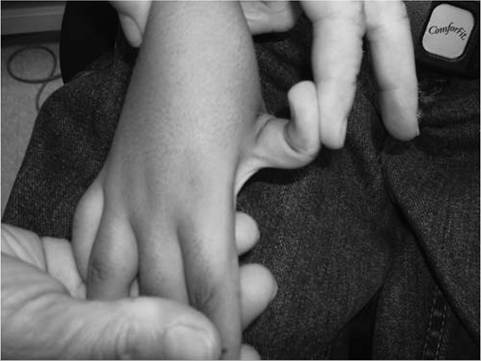
Figure 12.11 Joint laxity in Ullrich congenital muscular dystrophy with collagen VI abnormality.
VI expression may be absent in skeletal muscle and capillaries or absent on surface of muscle fibers but present in connective tissue. There has been no correlation between pattern of pathology and clinical phenotype.
Congenital Muscular Dystrophy With Early Spine Rigidity
This is a recessive condition caused by a defect in selenoprotein N, 1 (SEPN1) and linked to chromosome 1p35-p36.
Clinical severity is variable, with early-onset cases in infancy and later-onset cases in the later first decade. Patients present with hypotonia and poor head control. The weakness is symmetric and involves the neck, face, and proximal and distal musculature.Respiratory function is compromised with vital capacity below 55% by the end of the first decade. Patients often show signs of nocturnal hypoventilation and central apnea. Respiratory failure may develop. Some patients never develop walking. Muscle size is small, especially in the inner thighs and calves. Many children show early improvement, with development followed by nonprogressive or slow decline. The rigid spine develops by 3 to 7 years and is manifested by limited flexion of the neck and spine. Progressive scoliosis occurs with onset 4 to 12 years. Contractures of the elbow flexors, hip extensors, ankles, and knees are common. The rate of insulin resistance is increased, and intelligence is normal. Serum CK is usually normal. The muscle to biopsy can be best identified by MR imaging, with involved muscles often being the vastus lateralis and biceps femoris. Clinically, there is overlap with minicore congenital myopathy syndromes, and mutations in this SEPN1 gene also cause minicore congenital myopathy, congenital myopathy with desmin inclusions, and congenital fiber type size disproportion (small type I fibers).