Fascioscapulohumeral Muscular Dystrophy (FSHD)
Facioscapulohumeral muscular dystrophy (FSHD) is a slowly progressive dystrophic myopathy with predominant involvement of facial and shoulder girdle musculature (66). The condition has autosomal-dominant inheritance, with linkage to the chromosome 4q35 locus.
Approximately 10% to 30% of cases are caused by sporadic mutations. FSHD is the third most common of the dystrophies, behind Duchenne and myotonic dystrophies, with an incidence of between 10 and 20 per million live births (7). Age of presentation is generally before age 20. Changes on muscle biopsy are relatively slight, with the most consistent finding being the presence of isolated small atrophic fibers. Other fibers may be hypertrophied. Serum creatine kinase levels are normal or slightly elevated in the majority of patients. Diagnosis is confirmed in more than 90% of cases by molecular genetic testing.Facial weakness is an important clinical feature of FSHD muscular dystrophy. The initial weakness affects the facial muscles, especially the orbicularis oculi, zygomaticus, and orbicularis oris. These patients often have difficulty with eye closure but not ptosis. An individual may assume an expressionless appearance and exhibit difficulty whistling, pursing the lips, drinking through a straw, or smiling (Fig. 12.12). Even in the very early stages, forced closure of the eyelids can be easily overcome by the examiner. Masseter, temporalis, extraocular, and pharyngeal muscles characteristically are spared in FSHD.
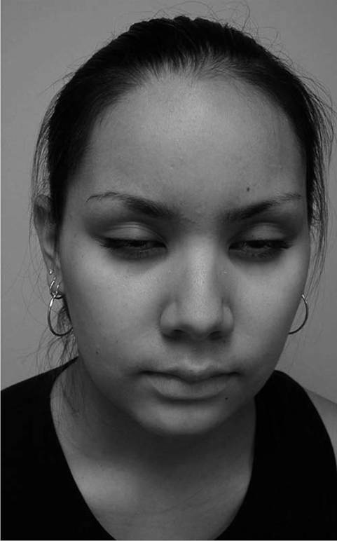
Figure 12.12 Facial weakness and expressionless facies in fascioscapulohumeral muscular dystrophy. Both father and daughter demonstrate difficulty whistling and pursing their lips.
Scapular stabilizers, shoulder abductors, and shoulder external rotators may be significantly affected, but at times the deltoids are surprisingly spared if tested with the scapulae stabilized.
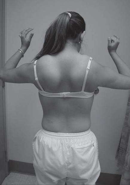
Both the biceps and triceps may be more affected than the deltoids. Patients with FSHD show characteristic patterns of muscle atrophy and scapular displacement. Involvement of the latissimus dorsi, lower trapezius, rhomboids, and serratus anterior results in a characteristic appearance of the shoulders, with the scapula positioned more laterally and superiorly, giving the shoulders a forward-sloped appearance (Fig. 12.13). The upper border of the scapula rises into the trapezius, falsely giving it a hypertrophied appearance. From the posterior view, the medial border of the scapula may exhibit profound posterior and lateral winging. The involvement of shoulder girdle musculature may be quite asymmetric. Some authors have found asymmetric weakness in the dominant upper extremity (67).A sensory neural hearing deficit was originally observed in Coates syndrome (early-onset FSHD). These individuals have a myopathy that presents in infancy. The disease progression is fairly rapid, with most individuals becoming wheelchair-reliant by the late second or third decade. These individuals also have a progressive exudative telangiectasia of the retina. Early recognition and photocoagulation of the abnormal retinal vessels may prevent loss of vision. Several audiometry studies have demonstrated hearing deficits in many later-onset FSHD patients in addition to those with Coates syndrome, suggesting that impaired hearing function is more common than expected in FSHD muscular dystrophy (68). Thus, all patients with FSHD should have screening audiometry and ophthalmologic evaluation. Contractures are relatively uncommon in FSHD muscular dystrophy. FSHD patients with scoliosis have mild and nonprogressive curves. Rarely, severe and progressive hyperlordosis is associated with FSHD. The patient with severe hyperlordosis may utilize their lordotic posturing to compensate for hip extensor weakness.
Mild restrictive lung disease has been reported in nearly one-half of FSHD patients (66).
The expiratory muscles involved in respiration appear to be more affected than inspiratory muscles in FSHD (67). Patients rarely require nocturnal ventilatory support.The presence of cardiac abnormalities in FSHD muscular dystrophy is debated. While diverse ECG abnormalities have been noted, one study showed no abnormalities on ECG, chest radiography, Holter monitoring, and echocardiography (69). Nuclear scanning with thallium-201 has demonstrated diffuse defects consistent with diffuse fibrosis (32). Abnormalities in systolic time intervals on echocardiography and elevations in atrial natriuretic peptide are consistent with subclinical cardiomyopathy. Cardiac complications in FSHD muscular dystrophy are rare, and patients in general have normal longevity. There is usually no associated intellectual involvement in this dystrophic myopathy.
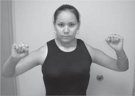
Figure 12.13 (A) Posterior and lateral scapular winging, high-riding scapula, and (B) hyperlordosis in Iascioscapulohumeral muscular dystrophy.