DISORDERS OF LYMPHATIC SYSTEM
The lymphatic system includes peripheral lymphocytes, lymphatic vessels and central lymphoid tissue, i.e. lymph nodes, spleen, tonsils, adenoids, Peyer's patches and thymus.
Lymph is an ultra-filtrate of blood (which oozed out into interstitial spaces) with a composition between transudate and exudate and contains variable number of lymphocytes.
It is typically milky in color (Chyle) due to presence of fat globules, except in early neonatal period before the feeding is established. From interstitial spaces, lymph is carried by tiny lymphatic capillaries (except in heart and brain) to drain into proximal lymphatic channels interspersed with regional lymph nodes, and finally to reach venous circulation directly or via thoracic duct that opens in left subclavian vein.Lymph nodes, consisting of well-defined dilated sinusoids lined by retiuloendothelial elements and surrounded by actively proliferating lymphocytes,
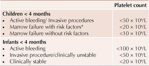
Fig. 19.14: Normal anatomy of cervical lymph nodes.
ALPS: Autoimmune lymphoproliferative syndrome
provide primary protective barrier against spread of regional infection. In sinusoids, lymph is filtered and particulate matters as well as living/dead organisms are phagocytised, processed and presented to further immune destruction.
According to location, lymph nodes are often termed as superficial (cervical, axillary, inguinal) or deeper (mediastinal, abdominal) groups.
Most frequently affected cervical group is divided into—(a) superficial horizontal chain or external Waldeyer ring, i.e. submental, submandibular, preauricular, post-auricular and suboccipital groups, (b) superficial vertical chain, i.e. anteromedial and posterolateral groups divided by sternocleidomastoid muscle (Fig.
19.14) and (c) deeper cervical lymph nodes.Some common disorders of lymph nodes or lymphatic channels are as follows:
Lymphadenopathy: Being the primary protective barrier against infection, lymph nodes are affected in many regional infections and systemic disorders.
Examination of superficial lymph nodes is an integral part of general examination and finding should be denoted in terms of: (a) size, (b) location or distribution, (c) consistency, (d) signs of inflammation, e.g. tenderness, overlying erythema, etc. (e) secondary changes, e.g. matting, fluctuation, sinus formation, scrofuloderma, etc. and (f) presence of any infective/inflammatory focus in draining area.
Deeper lymph nodes, i.e. mediastinal or abdominal lymph nodes may be assessed on chest skiagram and USG/CT respectively.
Etiology: Lymphadenopathy may be physiological or pathological (Table 19.34).
Pathological lymphadenopathy is indicated by—(a) size gt;1 cm (gt;1.5 cm in inguinal region), (b) involving multiple groups/regions, (c) firm/hard or fluctuating in consistency, (d) associated with secondary changes, and (e) presence of regional infective pathology.
Some typical features in common causes of lymphadenopathy in India are as follows:
• Physiological lymphadenopathy: Superficial lymph nodes may be normally palpable, specially between 4-8 years of age (i.e. period of normal lymphoid growth) and regress subsequently.
• Reactive lymphadenitis due to infective /inflammatory lesions in drainage area is localized, tender and transient, lasting for a few days. Streptococcal sore throat and viral pharyngitis are leading causes of unilateral and bilateral cervical lymphadenopathy, respectively.
• Tubercular lymphadenopathy is the commonest cause of persistent lymphadenopathy in Indian children, usually involving cervical or axillary nodes. Nodes are infected via lymphatic spread from intrathoracic nodes (natural infection) or vaccine site. BCG lymphadenitis is the commonest cause of axillary lymphadenitis in infancy (Ch 9.2).
Tubercular lymphadenopathy is characterized by firm, non-tender and matted nodes with/without secondary changes, e.g. sinus formation and scrofuloderma, i.e. exudative crusted skin lesions surrounding the sinus (Fig. 19.15).
• Neoplastic lymph nodes are typically non-tender, firm, fixed to skin/underlying structures, rapidly enlarging and/or involving multiple groups. In Hodgkin's disease, nodes typically have India-rubber consistency.
Diagnostic evaluation of lymphadenopathy is best achieved by fine needle aspiration cytology (FNAC) or lymph node biopsy, though supportive investigations, e.g. peripheral smear (for malignancy, infectious mononucleosis, etc.), tuberculin test and chest skiagram (for TB) and serology for other infections may clinch the diagnosis in many cases. In generalized
Fig. 19.15: Tubercular lymphadenopathy.
lymphadenopathy, it is also essential to evaluate status of deeper nodes by USG/CT scan.
Lymph node biopsy is indicated in: (a) clinically pathological lymph nodes, (b) presence of constitutional signs, e.g. fever, weight loss, etc., (c) increasing size for gt;2 weeks, and (d) no decrease/resolution in 6-8 weeks, despite antibiotics.
Lymphangitis is a regional inflammation of the lymphatic channels, presenting as tender-red streaks that extend proximally from infected site with enlarged and tender regional nodes. Staph. aureus and Group A #946; hemolytic streptococci are commonest organisms responsible for acute lymphangitis.
Lymphedema denotes obstruction of lymphatic flow, either due to congenital causes (Turner syndrome, Noonan syndrome, Milroy disease) or acquired pathology in regional lymph nodes, e.g. filariasis, neoplasms, postradiation or post-inflammatory scarring.
Cystic hygroma is a thin-walled, soft, fluctuant and brilliantly transilluminant cyst, filled with clear fluid, representing a lymphangioma or sequestered portion of embryonic jugular lymph sac.
These swelling are present at birth or manifest in early infancy, most common in posterior triangle of neck. Compression of neighboring vital structures or cyst infections are important complications of cystic hygroma.USG helps in antenatal and postnatal confirmation of cystic hygroma (multi-loculated echolucent mass). Treatment is surgical, with excision of as many cysts as possible while preserving vital structures, though laser therapy and sclerotherapy may be used for lesions not amenable to excision.
BIBLIOGRAPHY
1. World Health Organization. Haemoglobin concentrations for the diagnosis of anaemia and assessment of severity. Geneva: World Health Organization; 2011.
2. Chandra J et al. Diagnosis, Treatment and prevention of nutritional anemia in children: Recommendations of the Joint Committee of Pediatric Hematology Oncology Chapter and Pediatric and Adolescent Nutrition Society of the Indian Academy of Pediatrics. Indian Pediatr. 2022;59:782.
3. Bansal D et al. Iron deficiency anemia. Standard treatment guidelines. Indian Academy of Pediatrics. 2022
4. Dheenadayalan et al. Megaloblastic anemia. Standard treatment guidelines. Indian Academy of Pediatrics. 2022.
5. Rajaji S et al. G6PD Deficiency. Standard treatment guidelines. Indian Academy of Pediatrics. 2022.
6. Bafna V et al. Practicing pearls for sickle cell disease management. Standard treatment guidelines. Indian Academy of Pediatrics. 2022.
7. Indian Academy of Pediatrics: Thalassemia. Standard Treatment Guidelines. 2022.
8. Arya Y. Cell-based gene therapy for #946;-thalassemia. Indian Pediatr. 2023;.60.313.
9. Soni S. Gene therapies for transfusion-dependent #946;-thalassemia Indian Pediatr. 2021;58:667.
10. Danewa A et al. Diagnosis and management of acquired aplastic anemia: Consensus Statement of Indian Academy of Pediatrics. Indian Pediatr. 2022;59:467.
11. Indian Academy of Pediatrics: Immune Thrombocytopenia. Standard Treatment Guidelines 2022.
12. Sahi PK et al. American Society of Hematology Guidelines, 2019. Indian Pediatr. 202;57:854-56.
13. Neunert C et al. American Society of Hematology 2019 guidelines of IPB. Blood Adv. 2019;3:3829-66.
14. Rani S et al. Hemophilia. Standard treatment guidelines. Indian Academy of Pediatrics. 2022.
15. Sachdeva A et al. Consensus statement of the Indian Academy of Pediatrics in diagnosis and management of hemophilia. Indian Pediatr. 2018;55(7):582.
16. Srivastava A et al. WFH guidelines for the management of hemophilia, 3rd edition. Haemophilia. 2020:26(S 6):1-158.
17. Weyand AC et al. New therapies for hemophilia. Blood. 2019;133(5):389.
18. Helen V. Guidelines on transfusion for fetuses, neonates and older children. British Journal of Hematology 2016. 175: 784. Available on: https://onlinelibrary.wiley.com/doi/ full/10.1111/bjh.14233.