ECZEMATOUS SKIN DISORDERS
Eczema is a generic term to denote many hypersensitive skin disorders of heterogeneous etiology (Table 25.10), all characterized by:
• Typical weeping or oozing skin lesions, and
• Chronic course with intermittent exacerbations.
During acute phase, lesions are intensely pruritic and erythematous with microvesicle formation, while chronic lesions are typically dry and scaly with coarse skin markings (Iichenification) and altered pigmentation. Some important eczematous disorders are as follows: Atopic dermatitis is the commonest eczematous disorder in children with intermittent exacerbations and remissions, related to IgE-mediated cutaneous hypersensitivity.
Pathogenesis: Three important factors in causation or progression of atopic dermatitis are:
a. Intrinsic hyperreactivity of the skin, as evident by family history of atopy or white dermatographism, i.e. wheal and flare response on stroking the skin.
b. IgE-mediated allergic response, with intense pruritis in acute phase, followed by chronic inflammatory reaction. Introduction of a food-allergen, e.g. egg in early infancy has been implicated as a risk factor for sensitization.
c. Secondary infections during acute stage and licheni- fication of lesions during remission.
Clinical manifestations vary according to the age and phase of the disease. A typical case is characterized by:
a. Onset of skin lesions at 2-3 months of age, coinciding with introduction of certain foods in diet, e.g. animal milk, eggs, etc.
b. Intensely pruritic, erythematous weepy patches on cheeks, face, neck and extensor surface of extremities. Secondary infection is common (Fig. 25.15).
TABLE 25.10: Causes of eczema
• Atopic dermatitis
• Contact/irritant dermatitis
• Seborrheic dermatitis
• Pityriasis alba
• Nummular eczema
• Dyshidrotic eczema
c. Resolution of skin lesions by 1-2 years of age, followed by intermittent exacerbations and remissions.
Children with recurrent lesions develop lichenification, hyperpigmentation and scaling of earlier lesions with a whitish-hue on the face due to edema and blanching (mask-like face) and accentuated lines under the eyelids (Atopic pleat, Dennie's lines, Morgan's fold).
In older children, flexural regions, dorsum of hands/ feet, eyelids and neck are more frequently affected. In some, lesions remain localized as nummular eczema, i.e. coin-shaped, pruritic vesicular lesions (Fig. 25.16). Lasting remission usually occurs after fourth decade of life.
Diagnosis is largely clinical (Hanifin and Rajka criteria), based on presence of at least three of the following major criteria:
1. Typical morphology and distribution of lesions,
2. Presence of Pruritis,
3. Chronic or relapsing course, and
4. Family or personal history of atopy;
and, supported by any three of the many of the minor features listed below:
• Early age of onset (below 2 years),
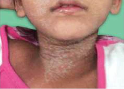
Fig. 25.16: Nummular eczema.
• Skin involvement with any of the following-xerosis, ichthyosis, palmar hyperlinearity, pityriasis alba, white dermatographism with delayed blanching, perifollicular accentuation, non-specific hand/foot dermatitis, Tendency for staphylococcal/HSV skin infections, Nipple eczema, Facial erythema/pallor, Cheilitis and Dennie-Morgan infraorbital folds;
• Accentuation of itching when sweating,
• Ocular lesions, e.g. recurrent conjunctivitis, anterior subcapsular cataracts, keratoconus,
• Immediate skin-test reactivity or elevated serum IgE levels
D/D of atopic dermatitis includes: (a) other causes of eczema (Table 25.10), (b) scabies, and (c) skin manifestations of systemic diseases, e.g. acrodermatitis enteropathica, histiocytosis, Wiskott-Aldrich syndrome, phenylketonuria, etc.
Management depends on the clinical phase:
• Acute phase with intense pruritis and inflammation is treated with topical wet dressings (Burrow's solution), antibiotics, e.g.
mupirocin and low-potency steroids, along with oral antihistaminics. Systemic steroids should be avoided, though systemic antibiotics may be required in presence of secondary infections.• During remissions, management aims to prevent dryness of skin by regular baths with lukewarm water for 5-10 minutes followed by immediate application of bland moisturizers, e.g. oil, creams, etc. after bath, to seal the skin moisture. Potential triggering factors,
e. g. frequent hand wash with soaps or chemicals, use of acrylic cloths and exposure to house dust mite must be avoided. Dietary restrictions are unlikely to be useful and Vaccinations are not contraindicated.
• Chronic Iichenified lesions may be managed with frequent (4-6 times) application of emollients after a lukewarm bath, with/without a short course of topical low-potency steroids or immunomodulators, e.g. tacrolimus, for significant pruritis. Antihistamines may also be used to break itching-scratch cycle. Narrow-band ultraviolet radiation or systemic immunosuppressive therapy may be rarely needed in extensive or resistant cases.
Contact dermatitis is a chronic skin reaction to various contactants, which may be immunological (Allergic contact dermatitis) or non-immunological (Irritant contact dermatitis) in origin.
Allergic contact dermatitis is a T-cell mediated hypersensitivity reaction (d/d IgE mediated atopic dermatitis), provoked by even transient contact (d/d irritant contact dermatitis) with an antigen or hapten. As initial sensitization takes few days, dermatitis develops only after persistent or re-exposure to same allergen.
Common examples of allergic contact dermatitis include: (a) ear-lobe dermatitis to metal earrings, (b) rhus
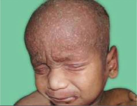
Fig. 25.17: Irritant contact dermatitis: (A) Diaper dermatitis; (B) Saliva dermatitis.
dermatitis to plant, e.g. poison ivy, congress-grass, etc,
(c) apparel-dermatitis to fabric dyes, fibers and elastics, and (d) cosmetic-dermatitis to powders, lipstick and nail polish.
Clinically, allergic contact dermatitis is characterized by erythematous, intensely pruritic weepy lesion, usually localized at the site of contact. Persistent/recurrent contact leads to development of typical eczema.
Management includes: (a) elimination of offending agent, (b) treatment of acute lesions with cool compresses, topical steroids and oral antihistaminics, (c) systemic steroids in severe reactions for 1-2 weeks, and (d) systemic antibiotics in secondary infections. Desensitization is not very effective in children.
Irritant contact dermatitis is caused by prolonged contact with endogenous irritants, e.g. saliva (over lips in habitual lip smackers), sweat (over feet after wearing tight shoes), urine (diaper dermatitis) or exogenous irritants (soaps, etc.)
Clinically, these lesions are characteristically localized and well-demarcated at the site of contact and usually begin as confluent erythema before turning into eczematous lesions over a period of time. Presence of red-satellite pustules indicates superadded candidal infection (Fig. 25.17).
Management includes: (a) avoidance of possible contact, e.g. wet diapers, (b) topical application of barrier agents, e.g. petroleum jelly/zinc oxide paste after cold compresses, and (c) topical short-acting steroids (1% hydrocortisone) in selected cases, though they increase risk of superadded candidal infection.
Dyshidrotic eczema is common in summer-season, characterized by recurrent crops of intensely pruritic, non-inflammatory small vesicles, over palm, soles and lateral aspects of fingers. Secondary infection is common due to scratching. Chronic lesions appear as thick, fissured plaques.
Exact etiology is unknown, though many cases have family history of atopy or hyperhidrosis. Treatment includes wet dressings and topical steroids in acute phase and topical steroids with a lubricant and keratolytic agent during chronic phase. Hands should be protected from chemicals, harsh soaps and exposure to adverse weather.
25.9