PAPULOSQUAMOUS DISORDERS
Papulosquamous disorders are characterized by presence of scales over the underlying plaque of patch. Some important papulosquamous disorders are as follows:
Psoriasis is a chronic autoimmune papulosquamous disorder, with ~1#8725;3rd cases manifesting in childhood.
Pediatric cases are more common in females and ~50% have positive family history.Etiopathogenesis is multifactorial. Trauma and drugs are known to precipitate/worsen skin lesions, in genetically predisposed individuals. Association with some HLA types (HLA-CW6) is known.
Clinically, these cases are characterized by chronic relapsing-remitting course with:
a. Erythematous papules/plaques with sharp demarcated borders, changing into thick silvery-white scales. Lesions are common over scalp, knees, elbows and lower trunk, often with symmetrical distribution (Fig. 25.13).
b. Positive Auspitz sign, i.e. pinpoint bleeding on removal of scales.
c. Positive Koebner response, i.e. appearance of new lesions at the site of trauma, e.g. scratching.
Clinically, these cases present with three stages:
• First stage manifests in newborns with typically linear and erythematous streaks/plaques of vesicles on limbs and trunk. Lesions usually resolve by 3-4 months, leaving dry, hyperkeratotic verrucous plaques for next 4-6 months. Intermittent recurrence during febrile episodes is common. Eosinophilia is present in gt;50% cases.
• Second stage (hyperpigmentary stage) is the hallmark of disease, characterized by development of hyperpigmented whorls, linear streaks or patches over trunk, axilla and groin. These lesions follow or overlap the first stage and persist till adolescence before fading.
• Third stage (hypopigmentary stage) with hairless, hypopigmented, anhidrotic patches/ streaks especially
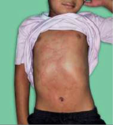
Fig.
25.13: Psoriasis.Based on morphology of lesions, psoriasis is classified as chronic plaque psoriasis, guttate psoriasis ands pustular psoriasis.
Diagnosis is clinical though needs to be differentiated from other papulosquamous disorders, e.g. pityriasis rosea, seborrhoeic dermatitis and lichen nitidus/ striatus/planus.
Management is mainly palliative, including:
• Avoidance of skin trauma.
• Topical steroids for localized plaques in early disease.
• Local application of 5-10% coal tar with/without ultraviolet or natural sun-light exposure is the treatment of choice for stable but established lesions.
Local keratolytic agents (1-3%) or topical Calcipotriene (a vitamin D analogue) may also be used.
• PUVA therapy with oral/topical psoralen and ultra- violet-A radiation for severe disease is effective but not safe in children. Other options for severe disease include methotrexate, cyclosporine and oral retinoids (Acitretin) therapy.
Seborrheic dermatitis in childhood is most common during infancy or adolescence. It is a chronic inflammatory disease of unknown etiology, though excessive perspiration, sebaceous gland dysfunction and infection with M. furfur have been implicated in etiology.
Clinical picture is variable, characterized by a relapsingremitting course. Re-activation is common during stress or due to poor hygiene.
Infants tend to have more severe and widespread disease with: (a) diffuse brawny scaling (cradle cap) or localized thick oily, yellowish crusts over scalp, occasionally involving eyelids and eyebrows, and
(b) greasy, scaly, erythematous and non-pruritic popular dermatitis over face, neck, behind the ears, axilla and groin, with post-inflammatory dyspigmentation (Fig. 25.14).
Fig. 25.14: Seborrheic dermatitis.
In adolescents, lesions are usually limited to scalp, intertriginous area and external auditory canal.
In severe cases, seborrheic plaques may be eczematous and intensely erythematous.Management includes: (a) regular use of anti-seborrheic shampoo (selenium sulfide), (b) topical steroids over acute lesion, and (c) topical antifungal ointment for M. furfur in some cases.
Pityriasis rosea is a common, benign papulosquamous disorder of probable viral in origin, seen mainly in young children.
Clinically most cases present after a preceding viral prodrome, with:
• A herald (mother) patch, i.e. solitary large (1-10 cm) round/oval lesion with raised border and fine adherent scales, followed after 5-10 days by
• Widespread symmetrical small (lt;1 cm) eruptions in crops, beginning from trunk and proximal extremities, which are raised, pink/brown in color and covered with a fine scale. Long axis of lesions corresponds with skin-cleavage lines, producing typical Christmas tree pattern.
• Self-limiting course with resolution in 2-6 weeks and no recurrence.
Treatment is not necessary for asymptomatic cases, though a bland emollient/lubricant lotion or topical steroids and antihistaminics may be used for itching.
Lichen planus is a common chronic dermatosis in adults, but rare in children, who account for lt;5% cases.
Etiologically it is considered as a T-cell mediated autoimmune disorder against unknown skin proteins. Onset of eruptions has been associated with exposure to some drugs.
Clinically, primary lesion in lichen planus is an intensely pruritic, flat-topped, polygonal and violaceous papule with fine whitish reticulations (Wickham striae) over the surface. These lesions are more common on flexor aspect of wrists, knee, anterior shins and tip of the penis. Koebner phenomenon (lesions induced by scratching) is common.
Orogenital mucosal involvement with lacy white papules is present in ~50% cases, though nail involvement is rare in children. Other variants include bullous, annular, linear and hypertrophic lichen planus. Lesions usually resolve after 12-15 months, but chronic course with intermittent exacerbations is common in older cases.
Treatment includes the use of systemic antihistaminics for pruritis and topical/ systemic/ intralesional steroids. Extensive lesions may benefit with narrow-band ultraviolet B radiation.Lichen nitidus in more common in children and young adults, presenting with tiny 1-2 mm monomorphic hypopigmented or flesh-colored papules with a shiny surface over arms, chest, abdomen and penis, often with
linear arrangement. Spontaneous disappearance after ~1 year is common, though a mid-potent topical steroid or tracrolimus may be used to relieve pruritis.
Gianotti-Crosti syndrome is a rare papular acrodermatitis in early childhood (1-6 years), usually associated with Hepatitis B infection. Eruptions begin as acute, multiple, tiny, lichenoid papules, which later coalesce to form plaques. Lesions are more common on face, buttocks and extremities, typically sparing the torso. Apart from moderately severe pruritis, the children are generally well and lesions resolve spontaneously after 2-8 weeks.
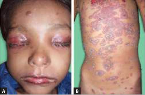
Fig. 25.15: Atopic dermatitis.
25.8