FILARIASIS
About 2/3rd of filaria cases in world are from three Asian countries, i.e. China, India and Indonesia. It is endemic
all over India, except in some northern and north-eastern states.
National Health Policy (2017) aims to eliminate filaria, defined as achieving microfilaria rate of lt;1:10000 population in endemic regions.Epidemiology: In India, 98% cases of filariasis are caused by W. bancrofti, and rest by a closely related Brugia malayi infection, limited to certain districts of Kerala.
Reservoir of infection is an infected man, harbouring adult worms of ~4-5 cm size in lymphatic system that live for many years. Female worm produces ~ 50,000 microfilaria (Mf) per day, which enter the circulation via lymphatics, usually at midnight (nocturnal periodicity).
Mode of transmission is a vector (Mosquito)-Culev fatigans for bancroftian filariasis and Mansonia annulifera for brugian infection. After an infected feed, Mf exsheath in mosquito gut to develop as infective larva (10-14 days), which migrate to proboscis and injected into new human host during next bite.
After the infected bite in humans, infective larva are deposited near puncture-site and enter lymphatics, where they reside and develop as adult worm to produce Mf.
Clinical manifestations develop only in a small percentage of infected population, less common in children than adults and present as: (a) lymphatic filariasis, or (b) occult filariasis.
• Lymphatic filariasis may present as:
- Asymptomatic microfilaremia, detected on nightsmear examinations in endemic zones,
- Acute filariasis in initial months of infection, with recurrent episodes of fever, lymphangitis, lymphadenitis, lymphedema and epididymo-orchitis, and
- Chronic lymphatic obstruction due to fibrosis and block of lymphatic channels after many years, e.g. elephantiasis, hydrocele and chyluria (very rare in childhood).
• Occultfilariasis, without signs of lymphatic disease, is more common in children and presents with tropical pulmonary eosinophilia or other immunological disturbances, e.g. arthritis, proteinuria, etc.
Diagnosis usually depends on:
• Clinical features of acute/obstructive disease in endemic area, supported by,
• Night-smear: Demonstration of Mf in thick peripheral smear (Fig. 10.21), collected around mid-night or after one hour of 100 mg PO diethylcarbamazine administration (Provocation test);
• Serodiagnosis i.e demonstration of filarial antigens/ antibodies, and
• Biopsy/radiology of affected part showing calcified, dead or living worm/s.
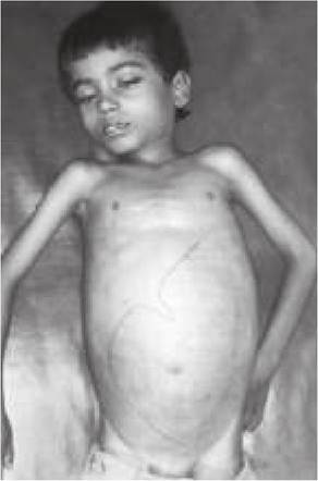
Fig. 10.21: Microfilaria: Peripheral smear.
TABLE 10.58: Diagnostic criteria for TPE
• Typical clinical episodes in endemic region
• Hypersensitivity indicators:
- Absolute eosinophil count gt;3000 cells/mm3
- Elevated IgE levels (gt;1000 IU/ml)
- Presence of anti-Mf antibodies
• Therapeutic response to DEC therapy
Treatment: Role of anti-filarial drugs in acute filariasis or chronic obstructive disease is controversial, as these drugs have no effect of infective larvae or ? adult worms. However, all Mf positive cases need specific therapy to reduce parasite load, as follows:
• PO diethylcarbamazine or DEC (6 mg/kg/d q8hr) for 14-21 days. Some workers recommend lower dose on first 2-3 days to avoid hypersensitivity reactions due to rapid death of Mf.
• PO Ivermectin (20-200 mg/kg) with/without PO albendazole (400 mg), as a single dose is equally effective to clear microfilaremia.
Tropical pulmonary eosinophilia is a common hypersensitivity manifestation against Mf or ? adult worms in school children of endemic regions.
Clinically these cases present with recurrent and paroxysmal asthma-like episodes of dry cough, wheeze and dyspnea; with/without mild fever, anorexia, hepatosplenomegaly and lymphadenopathy. Recurrent episodes may lead to interstitial fibrosis and chronic respiratory insufficiency.
Diagnosis rests on a diagnostic criteria (Table 10.58) among residents of an endemic region. Night smears for Mf may/may not be positive. X-ray chest may be normal except bronchovesicular prominence or reveals typical miliary mottling.
D/D of TPE includes other causes of paroxysmal cough with eosinophilia, e.g. asthma, Loeffler syndrome, drug allergy, fungal infections, sarcoidosis, lymphomas and rare hypereosinophilic syndromes.
Treatment involves oral DEC therapy for 21 days, which may be repeated in cases with partial response. Response is monitored clinically and by absolute eosinophil counts.
10.36