LIVER ABSCESS
Pyogenic liver abscesses are not uncommon in children, Solitary liver abscesses in the right lobe are more common than multiple abscesses or solitary left lobe abscesses.
Etiologically, liver abscesses usually develop as a part of generalized sepsis, as ascending infection from gastrointestinal disease or localized lesions after intraabdominal infection, trauma or surgery.
Most common pathogenic organisms include Staphylococcus aureus, Streptococcus spp., Escherichia coli, Salmonella, and anaerobic organisms. Entamoeba histolytica is an important cause of liver abscess in India, though more common in adults.
Clinically these children present with fever, chills, vomiting and abdominal pain in right hypochondrium with tender hepatomegaly. Jaundice is uncommon.
Complications include spontaneous rupture in peritoneum, pericardium or pleural space or metastatic abscesses at other sites.
Diagnosis rests on USG/CT followed by guided percutaneous aspiration to confirm the microbial etiology (Fig. 15.2). Presence of elevated right hemidiaphragm with leukocytosis indicates possibility of liver abscess in a clinically suspected case.
Treatment: Drainage and appropriate antibiotics are mainstays of treatment in liver abscess.
Initial antibiotic regimen should be directed against gram-positive aerobes, gram-negative bacilli, and
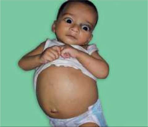
Fig. 15.2: Liver abscess.
anaerobic organisms with subsequent modification according to culture reports for a total duration of 4-6 weeks. Serial USG may be required to assess the response to therapy.
Percutaneous drainage is indicated where lesions are accessible under CT or ultrasound guidance. Catheters placed via these techniques are generally left in place until abscess collapse (pig tail).
Open surgical drainage may be required if percutaneous drainage is not feasible or fails.TABLE 15.10: Causes of chronic liver disease
• Infective
- Hepatotropic virus infections: HBV, HCV
- Intrauterine (TORCH) infections
• Metabolic liver disease
- Common: Wilson disease, Gaucher disease, GSD
- Rare: Cystic fibrosis, Indian childhood cirrhosis
- Others: Galactosemia, #945;1-antitrypsin deficiency
• Drug/toxic liver injury
- Veno-occlusive disease
- Drugs: Methotrexate, INH, etc.
• Autoimmune liver disease
• Neonatal cholestasic syndromes (Table 12.41)
GSD: Glycogen storage disorders
Depending on the nodular size, cirrhosis is also termed
(a) micronodular, e.g. Indian childhood cirrhosis, biliary cirrhosis, etc.; (b) macronodular, e.g. Wilson disease, post necrotic/hepatitic cirrhosis, etc. or (c) mixed, e.g. metabolic liver diseases, etc.
Clinical spectrum of CLD ranges from—(a) asymptomatic biochemical abnormalities, to (b) signs of progressive liver disease, or (c) signs of acute decompensation over pre-existing liver disease. Liver may or may not be palpable, depending on primary pathology. Important clinical features of CLD are tabulated in Table 15.11.
More on the topic LIVER ABSCESS:
- Hepatic Lobe Torsion
- Liver Transplantation
- Liver Disease
- LUNG ABSCESS
- HEPATOMEGALY
- Arcanobacterium infections
- ABDOMINAL MASS/LUMP
- Agrawal M.. Textbook of Pediatrics. 3rd ed. — CBS Publishers,2025. — 973 p., 2025