PATHOLOGY
More than 80% of children with CP will have abnormal findings on neuroimaging (33-35). These abnormal findings can provide valuable clues to pathogenesis.
The most common abnormality on neuroimaging is found in the white matter near the lateral ventricles, often termed periventricular leukomalacia (PVL), with reports of up to 56% of all cases of CP demonstrating abnormalities in this location (34) (Fig.
8.7). PVL occurs much more commonly in premature infants than in term infants (90% vs 20%) and is a common outcome of intraventricular hemorrhage in premature infants (34). Because the corticospinal tract fibers to the lower extremities are medial to those of the upper extremities in the periventricular white matter, children with PVL typically have spastic diparesis. One large study found that PVL was present in 71% of the children with diparesis, 34% of those with hemiparesis, and 35% of those with quadriparesis (33).Deep grey matter lesions to the basal ganglia and thalamic region are mainly associated with dystonic CP, and have been found in approximately 12% of children
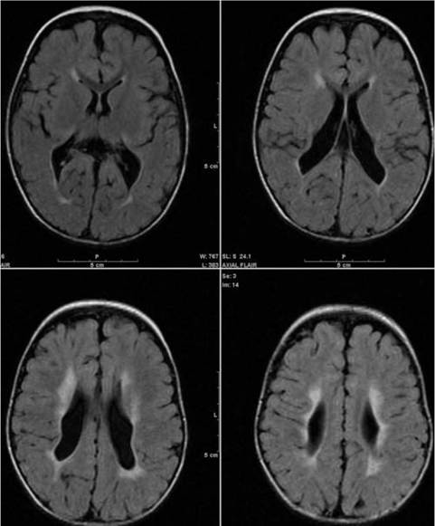
Figure 8.7 Periventricular leukomalacia.
with the condition (33). Historically, large numbers of children acquired athetoid CP following a diagnosis of kernicterus, due to concentrated damage to the basal ganglia with bilirubin encephalopathy. These cases are far less common with advancements in the treatment of neonatal jaundice.
Focal cortical infarcts involving both the grey and white matter are found almost exclusively in patients with hemiparesis, and are typically related to middle cerebral artery strokes. In a group of children with hemiparetic CP, 27% were found to have a focal infarct on imaging (33).
Brain malformations can be found on neuroimaging in approximately 10% of children with CP (33-35). Neuronal migrational disorders early in pregnancy can result in lissencephaly, polymicrogyria, schizen- cephaly, or holoprosencephaly. Some in utero infections, such as those caused by cytomegalovirus, can also cause distinctive brain malformations (33). Brain malformations are more commonly found in cases of term infants and hemiparesis (35).
Children who sustain diffuse brain insults demonstrate more extensive injury on neuroimaging. Infection and ischemia are two of the more common causes of generalized encephalomalacia. A wide range of findings may be present on magnetic resonance imaging (MRI), including multiple cysts, cortical thinning, white and grey matter loss, and microcephaly. Children with diffuse brain lesions or anomalies typically demonstrate spastic quadriparesis and are at high risk for additional medical and cognitive problems.