PENOSCROTAL DISORDERS
Penoscrotal disorders may be congenital or acquired. This chapter deals with some important penosrcotal problem in children excluding penile malformations, i.e. epispadias/hypospadias, discussed earlier (Ch 21.13) and urogenital infections, which are more frequent in adolescents and discussed in Ch 13.3.
Cryptorchidism (undescended testis): Testes in early fetal life develop intra-abdominally and descend subsequently into scrotum, guided by its anchor - Gubernaculum. Testicular descent is controlled by various mechanical factors, i.e. intra-abdominal pressure, gubernacular-pull, etc. as well as hormonal factors, i.e. testosterone, dihydrotestosterone and Mullerian- inhibiting factor.
Cryptorchidism (absence of testes in scrotum) represents complete/partial failure of normal testicular descent, arrested at any point along the normal path, i.e. abdomen (15%), inguinal canal (80-85%) or rarely migrated to ectopic sites, e.g. perineum (lt;5%).
Incidence: Cryptorchidism is present in ~4-5% of term newborns and in lt;1% at 1 year of age. As the testicular descent mainly occurs in last trimester, incidence is naturally higher in preterms.
Etiology: Various factors, implicated in the etiology of undescended testis include—(a) mechanical factors, e.g. Inguinal hernia, abnormal gubernaculum; (b) hormonal factors, e.g. androgen deficiency or insensitivity, and (c) abnormalities of epididymis or genitofemoral nerve.
Clinically, cryptorchidism is usually unilateral (80-90%). True undescended testis must be differentiated from retractile testis, i.e. testis palpable in inguinal canal, but can be pulled down into scrotum. Retractile testis is not uncommon due to overactive cremasteric reflex in older boys and hence, should be examined in squatting position. Indirect inguinal hernia is common with cryptorchidism (Fig. 21.10).
Complications: Undescended testis is essentially normal at birth but develops progressive pathological changes, due to higher temperature and other factors operative at ectopic sites.
Common complications include:• Infertility, more common in bilateral (~40-50%) than unilateral (~10-15%) cryptorchidism. However, children operated before 2 years usually retain normal testicular functions.
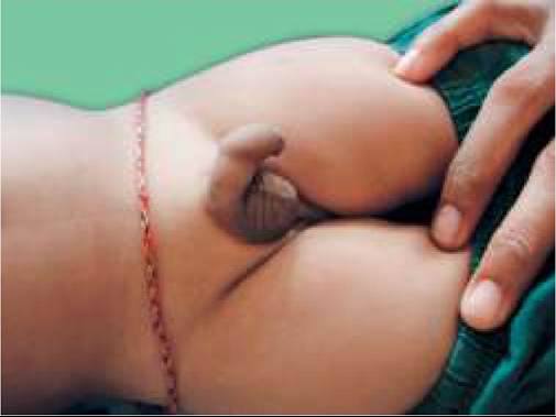
Fig. 21.10A and B: Cryptorchidism: (A) Bilateral; (B) Unilateral.
• Testicular malignancy in 3rd#8725;4th decade is ~10 times more common in undescending testis.
• Testicular torsion with/without infarction.
Diagnosis is largely clinical, though investigations are needed to—(a) locate abdominal testis by USG, MRI or laparoscopy, and (b) to exclude anorchia (absent testis), by hormonal tests, e.g. FSH, LH and testosterone levels after HCG stimulation.
Treatment: As ~80% of undescended testis, specially those in inguinal canal, descend spontaneously in infancy, surgery is deferred till 9-15 months. However, further delay is not advisable due to risk of irreversible pathological changes, which may affect fertility.
Orchidopexy with closure of indirect hernia is the treatment of choice in most cases. Two-stage orchiopexy may be needed for high abdominal testis. Hormonal treatment with human chorionic gonadotropin (hCG) or gonadotropin releasing hormone (GnRH) may be tried to facilitate testicular descent in cases with low/retractile testes with limited success.
Orchiectomy is indicated in children with delayed diagnosis (gt;2 years) and non-functioning testis, to reduce the risk of malignancy in later life. Buserelin (a GnRH analogue) may be used in selected cases with delayed orchidectomy, to normalized testicular histopathology, though with variable results.
Hydrocele, i.e. accumulation of fluid in the tunica vaginalis, may be congenital or acquired.
Etiology: Most cases of childhood hydrocele are congenital due to persistent patency of processus vaginalis, which may/may not be communicating with abdomen at the time of diagnosis.
Acquired hydrocele, although uncommon, may be due to testicular torsion, epididymitis, trauma or tumors. Filaria-a common cause of hydrocele in Indian adults, is uncommon in childhood. Clinically, hydrocele presents as a smooth, non-tender, trans-illuminating scrotal swelling. Communicating hydrocele is often associated with indirect inguinal hernia and reducible on pressure (Fig. 21.11).
Fig. 21.11: Congenital hydrocele.
Management: Non-communicating hydrocele usually absorbs spontaneously by 12 months of age and Surgical drainage is indicated only in—(a) hydrocele persisting gt;12 months, (b) large/tense swellings, and (c) acquired hydrocele. Congenital communicating hydrocele requires ligation of processus vaginalis with herniorrhaphy.
Varicocele, i.e. abnormal dilatation of pampiniform venous plexus of scrotum due to valvular incompetence of spermatic vein is rare before adolescence and should raise a possibility of retro-peritoneal mass.
These cases present with painless para-testicular mass with 'bag of worms' feel on palpation which becomes prominent on erect position.
Varicocelectomy is indicated only in children with large varicocele or significant disparity in testicular size. Micropenis is defined as the normally formed penis, but with stretched penile length of lt; 2 SD below the mean size for age (lt; 1.9 cm in newborns).
It is caused by hormonal deficiency after the first trimester, due to: (a) hypogonadotropic hypogonadism, e.g. in Lawrence-Moon-Biedl syndrome, Prader-Willi syndrome, etc. (b) hypergonadotropic hypogonadism, i.e. primary testicular failure, and (c) idiopathic micropenis.
Evaluation includes karyotyping and endocrinal studies to assess pituitary and testicular functions. Androgenic hormonal therapy may be useful in some cases, though most cases achieve normal sexual function even without therapy.
Inflammatory penile disorders of penis or scrotum in children are generally infective or traumatic in origin.
Some important disorders include:Phimosis, i.e. inability to retract the prepuce, is physiological at birth, that corrects spontaneously by 3 years of age in gt;90% cases. In older boys, it may be: (a) congenital, (b) secondary to prepuceal infection/ inflammation with scarring, or (c) post-circumcision. True phimosis is uncommon and occurs due to recurrent balanoposthitis causing fibrous rings.
No treatment is needed for physiological phimosis, except frequent cleaning of sub-prepuceal debris. In older children, local application of a steroid cream for 1-2 months may loosen the phimotic ring sufficiently to enable prepuceal retraction.
Circumcision on medical grounds is recommended only in—(a) phimosis persisting beyond 10 years or (b) ballooning of prepuce during micturation, and (c) associated paraphimosis.
Role of preventive circumcision in infancy to reduce the risk of—(a) secondary balanoposthitis and phimosis, (b) obstructive uropathy or UTI, (c) sexually transmitted diseases, e.g. HIV, and (d) penile carcinoma in later life, is highly controversial.
Common complications of circumcision include: (i) local bleeding, (ii) wound infection, (iii) secondary phimosis, and (iv) meatal stenosis. It is contraindicated in micro-penis, to spare extra skin for later reconstructive surgery.
Paraphimosis, i.e. retraction of prepuceal skin behind the coronal sulcus that can't be pulled back over glans, is a more serious problem, leading to painful venous stasis in the foreskin with edema. Treatment includes gentle stretching of prepuceal skin after lubrication, though emergency circumcision may be required.
Meatal stenosis is rarely primary, usually develops after circumcision due to inflammation of denuded glans. These cases present with dysuria and thin urinary stream with recurrent urethritis, urinary stasis and urinary tract infection.
Serial dilation of meatus is recommended as initial treatment, while resistant cases may need meatotomy or meatoplasty.
Scrotal swellings/masses may be acute or chronic, painless or painful (Table 21.31). Diagnosis is usually established by clinical evaluation, USG and testicular flow scan (99mTc-pertechnectate) to establish vascular flow (absent in torsion, increased in inflammatory conditions). Some important causes are as follows.
Testicular torsion of spermatic cord is the commonest cause of acute, painful, scrotal swelling in adolescents (rare lt;10 years), due to abnormal fixation of testis in the scrotum. Absent cremasteric reflex is strongly indicative of testicular torsion.
As the vascular flow to testis is rapidly interrupted in these cases, Immediate surgical de-torsion with refixation is essential within 6 hours, for survival of gonads.
Torsion of testicular appendix is the commonest cause of acute scrotum lt; 2 years, rare in adolescents (d/d torsion of spermatic cord). Testicular blood flow is usually maintained in this condition and spontaneous recovery with conservative management is common.
Epididymo-orchitis is the principle cause of acute painful scrotal swelling in adolescents, usually suggestive of sexually transmitted diseases, e.g. gonorrhoea or chlamydial infections. Epididymitis is rare before puberty and indicates possibility of an underlying Wolffian duct anomaly.
Testicular tumors may develop at any age including newborns. Nearly 2/3rd of them are malignant (germ cell tumors) and present as painless, hard testicular mass without transillumination.
Diagnosis rests on USG-guided FNAC or biopsy, along with serum tumor markers. Radical orchidectomy is essential in most cases.
BIBLIOGRAPHY
1. Uthap S et al. Acute glomerulonephritis. (Acute nephritic syndrome): Standard treatment guidelines. Indian Academy of Pediatrics 2022.
2. Bagga et al. Hemolytic uremic syndrome in a developing country: Consensus guidelines. Pediatric Nephrology 2019.
3. Bagga A et al. Nephrotic syndrome: Standard treatment guidelines. Indian Academy of Pediatrics 2022.
4. Sinha A et al. Steroid sensitive nephrotic syndrome: revised guidelines. Indian Pediatr. 2021:58:461.
5. Vasudevan A et al. Consensus guidelines on management of steroid-resistant nephrotic syndrome. Indian Pediatr. 2021:58:650.
6. Manthan M et al. Acute kidney injury: Standard treatment guidelines. Indian Academy of Pediatrics 2022.
7. Kidney disease: Improving global outcomes (KDIGO) Acute kidney injury group. Clinical practice guidelines for acute kidney injury. Kidney Inter. 2012; 2:1.
8. Ekambaram S et al. Urinary tract infection in children: Standard treatment guidelines. Indian Academy of Pediatrics 2022.
9. Shah M et al. Antenatally detected hydronephrosis standard treatment guidelines: Indian Academy of Pediatrics 2022.
10. Mane S et al. Undescended testes and testicular torsion: Standard treatment guidelines. Indian Academy of Pediatrics 2022.
11. Chandrasekharan VVS et al. Inguinal hernia and hydrocele: standard treatment guidelines. Indian Academy of Pediatrics 2022.