Posttraumatic Hydrocephalus and Cerebral Atrophy
Ventriculomegaly is seen commonly after severe TBI in children (61). The enlargement of the ventricular system can be either from high-pressure hydrocephalus or from cerebral atrophy resulting in hydrocephalus ex vacuo.
True hydrocephalus is a result of either an obstruction in the cerebral spinal fluid flow or impairment in the absorption of cerebral spinal fluid, ultimately resulting in an increase in cerebral spinal fluid volume and pressure. Hydrocephalus can be described, therefore, as either communicating (where there is abnormality in absorption) or noncommunicating (where there is an obstruction in the flow of the cerebral spinal fluid). The majority of hydrocephalus is caused by impaired cerebral spinal fluid absorption, secondary to inflammation or secondary to subarachnoid hemorrhage.Hydrocephalus ex vacuo describes enlargement of the ventricular system that results after cerebral atrophy and loss of brain volume (Fig. 10.1). To distinguish between clinically significant hydrocephalus and the expected consequence of cerebral atrophy after severe TBI, one must consider the patient's clinical status as well as the amount of time that has passed since the injury. Overall, if the patient is continuing
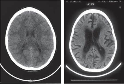
Figure 10.1 Cerebral atrophy. (A) Normal computed tomography (CT) scan. (B) CT scan showing posttraumatic brain injury cerebral atrophy with ventriculomegaly and increased sulci.
to demonstrate ongoing and regular improvements in their clinical status, Ventriculomegaly is more likely to be due to cerebral atrophy. The patient who has hydrocephalus typically continues with poor clinical improvement or clinical deterioration. The CT scan findings will yield clues as well, with cerebral atrophy demonstrating areas of encephalomalacia or enlargement of sulci, while hydrocephalus demonstrates more specific changes around the ventricular system outlined in Table 10.2 (203).
Hydrocephalus should be suspected if clinical improvement is not noted in a patient status post-TBI or if the clinical picture includes functional decline, seizures, abnormal posturing, or increased tone. Consideration of hydrocephalus in these patients is paramount, as failure to identify hydrocephalus when it is present may delay recovery. CT scan of the brain allows for rapid detection of hydrocephalus. The treating team may then choose to have a ventricular peritoneal shunt placed, which may improve the clinical status of the patient when normal ventricular pressures are reestablished (204).
More on the topic Posttraumatic Hydrocephalus and Cerebral Atrophy:
- Congenital/Hereditary Hydrocephalus
- Spinal Muscular Atrophy II
- Hydrocephalus
- TECHNICAL FACTORS OF NEEDLE ELECTROMYOGRAPHY