SWEAT GLAND DISORDERS
Sweat glands include two types of exocrine glands- apocrine glands and eccrine glands.
Eccrine glands, distributed all over skin including palm and soles, are prime regulators of body temperature.
These glands, located in dermis or subcutis, produce sweat that is secreted over skin surface via sweat pores.Apocrine glands are mainly located in axilla, areola, anogenital area and periumbilical region and continuously secrete an odorless milky fluid (apocrine sweat) during and after puberty. Bacterial decomposition of apocrine sweat is responsible for unpleasant odor of perspiration.
Miliaria rubra and M. crystallina is the commonest disorder of sweat glands due to a occlusion of their ducts by a keratin plug, discussed in Chapter 25.5.
Anhidrosis or Hypohidrosis, i.e. absent or less sweating over all or part of the skin may be due to:
a. Neural pathway disturbances, e.g. hypothalamic tumors, brain-stem lesions, spinal lesions (syringomyelia), peripheral neuropathies (leprosy), autonomic disorders (Riley-Day syndrome); or
b. Sweat gland abnormalities, e.g. congenital absence (ectodermal dysplasia), atrophy or destruction (burns, radiotherapy), drug-induced paralysis (atropine, scopolamine), blocked sweat ducts (miliaria, ichthyosis, atopic eczema).
Severe anhidrosis alters thermoregulatory mechanisms and may present with hyperthermia. Absence of sweating is a diagnostic feature of heat stroke and distinguishes it from heat exhaustion. Hypohidrosis is common in severe dehydration. Sweating is minimal in early infancy.
Hyperhidrosis, i.e. excessive sweating may be due to high environmental temperature, exercise or emotional disturbances, though pathological causes include: (a) neurological disorders, e.g. Riley-Day syndrome or spinal cord injuries, (b) metabolic states, e.g. hypoglycemia, hyperthyroidism, rickets, (c) drugs, e.g.
antipyretics, insulin, (d) congestive cardiac failure.Bromhidrosis, i.e. excessive odor of sweat, is a common problem in adolescence, due to formation of short-chain
fatty acids and ammonia by action of anaerobic diphtheroids on axillary apocrine sweat. Cleaning with a germicidal soap and topical application of aluminum/ zinc containing powders is often enough in most cases.
25.11.4 NAIL DISORDERS_________________________
Nails are specialized cuticular structures, consisting of nail plate bounded by lateral and posterior nail folds, covering a crescentic white area (Lunula) and underlying vascular bed (giving pink color to nails). Nail abnormalities may be primary or acquired after trauma, infection or chronic systemic diseases (Table 25.13).
Acute paronychia, i.e. infection of nail bed is usually caused by staphylococci or streptococci, and presents with inflammation and severe pain/tenderness of proximal nail bed (Fig. 25.18 A). Treatment includes oral antibiotics, e.g. amoxicillin/clavulanate, warm compresses, anti-inflammatory agents and incision/ drainage in some cases.
Chronic paronychia is more common in children with: (a) finger/thumb sucking, (b) embedded foreign body, e.g. wood splinter, (c) nail trauma, (d) chronic submersion of nails in irritating solutions, (child labor), and (e) diabetes.
| TABLE 25.13: Common nail abnormalities | |
| Nail abnormality | Cause |
| Deformity: | |
| Anonychia (absent nails) | Traumatic, congenital |
| Platynychia/Koilonychia | Iron deficiency anemia, hereditary, developmental* |
| Clubbing | See Chapter 17.2 |
| Hypoplastic nails | Nail-patella syndrome |
| Onycholysis (destruction) | Trauma—nail biting Infection—onchomycosis |
| Periungual fibroma | Tuberous sclerosis |
| Discoloration: | |
| Pallor | Anemia |
| Leukonychia (White lines) | PEM, hypoproteinemia Trauma, infection |
| Yellow | Meconium staining* Yellow-nail syndrome |
| Black pigmentation | Normal (10-15%) Chronic infection, traumatic |
| Splinter hemorrhage | Inferior endocarditis, trauma, Chronic liver disease |
*in infants
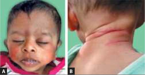
Fig. 25.18: (A) Acute paronychia; (B) Paronychia.
Microbially, it is often caused by candida or mixed bacterial flora and presents with persistent swelling of proximal nail fold, separation of nail plate and suppuration (Fig. 25.18B). Management includes keeping the nails dry and prolonged topical antibiotic/ antifungal therapy.
In-grown nails, i.e. penetration of lateral nail fold/ or spicule into surrounding soft tissue may lead to recurrent paronychia and usually seen in cases with poor-fitted footwear and improperly cut nails. Management includes: (a) treatment of local paronychia, (b) cauterization of soft granulation tissue over lateral nail bed, and (c) nail removal in recurrent cases. Nail should be allowed to grow beyond free edge before cutting transversely rather than in curvilinear fashion.
Nail-patella syndrome is an autosomal dominant disorder, characterized by hypoplastic nail/s and patella (knee instability) with/without cutis laxa, hyperextensible joints, hyperhidrosis and renal anomalies.
Yellow-nail syndrome (thickened, excessively curved, yellowish nails, without lunula) is seen in bronchiectesis and chronic lymphatic limb edema, e.g. filariasis.
BIBLIOGRAPHY
1. Madhu R et al. Indian Academy of Pediatrics Guidelines for Pediatric Skin Care. Indian Pediatr. 2021;58:1155.
2. Anandh Parchuri A, Saboth P. Tinea infections standard treatment guidelines. Indian Academy of Pediatrics. 2022.
3. Thomas J et al. Urticaria. Standard treatment guidelines. Indian Academy of Pediatrics. 2022.
4. Bhaskar V et al. Atopic dermatitis standard treatment guidelines. Indian Academy of Pediatrics. 2022.
5. Sarkar R et al. Steven Johnson syndrome/toxic epidermal nectrolysis standard treatment guidelines. Indian Academy of Pediatrics. 2022.
6. Rengasamy M et al. Staphylococcal scalded skin syndrome. Standard treatment guidelines. Indian Academy of Pediatrics. 2022.
More on the topic SWEAT GLAND DISORDERS:
- Zymbal's Gland and Preputial Gland Tumors
- Agrawal M.. Textbook of Pediatrics. 3rd ed. — CBS Publishers,2025. — 973 p., 2025