ANATOMIC PLANES OF REFERENCE
There are four anatomic planes of reference, two of which are variations of each other. Each plane is an imaginary slice through the body that is oriented at right angles to the other two.
The four reference planes (Figure 1-1) are as follows: Sagittal plane: A plane that runs the length of the body and divides it into left and right parts that are not necessarily equal halves.Median plane: A special kind of sagittal plane that runs down the center of the body lengthwise and divides it into equal left and right halves. It could also be called a mid- sagittal plane, but that term is not commonly used.
Transverse plane: A plane across the body that divides it into cranial (head-end) and caudal (tail-end) parts that are not necessarily equal.
Dorsal plane: A plane at right angles to the sagittal and transverse planes. It divides the body into dorsal (toward the animal’s back) and ventral (toward the belly) parts that are not necessarily equal. If an animal stands in water with its body partially submerged, the surface of the water describes a dorsal plane. In humans this plane is called the frontal plane (Figure 1-2).
CLINICAL APPLICATION
TEST YOURSELF 1-1
1. How does the anatomy of a muscle or bone differ from its physiology? Which describes appearance and location and which describes function?
2. How might abnormalities in an animal's anatomy or physiology have a negative impact on its health and well-being?
TERMINOLOGY
To be clear and accurate with descriptions of body parts, we have to use terms that leave no doubt about their meanings. Terms such as up, down, above, below, and beside are not very useful because they depend on the orientation of the animal (upright, on its side, on its back, and so on). If an animal is lying on its left side, is its right lung above its left lung or beside it? If the animal stands up, what is the relationship between the lungs then? Even the position of the observer can make a difference in terms such as left and right. If a structure in an animal is located “to the right” of another structure, does the meaning change if
Radiography Positioning Terminology
Radiographs, commonly called x-rays, are two-dimensional images of what is inside an animal.
Radiographs are described according to the path the x-ray beam takes through the body using anatomic directional terms. For example, imagine a dog lying on an x-ray table on its back. The x-ray tube is above it, and the x-ray film is beneath it in a light-tight case called a cassette. During the exposure, the x-rays will enter the animal’s ventral surface, pass through the abdomen, and exit the animal’s dorsal surface before striking the film. We call this a ventro-dorsal (VD) view of the abdomen, because the x-rays enter the ventral surface and exit the dorsal surface of the body. A dorso-palmar (DP) view of a horse’s front fetlock joint, which is the joint between the large metacarpal bone and the proximal phalanx, will have the x-ray machine positioned in front of the leg and the x-ray cassette behind the joint. The x-rays will enter the dorsal surface of the leg and exit the palmar surface. Lateral radiographic views are taken by passing the x-ray beam through the area of study from side to side. They are named according to which side of the animal is closest to the film. If the animal’s right side is closest to the film for an abdominal radiograph, the view is called a right lateral view of the abdomen.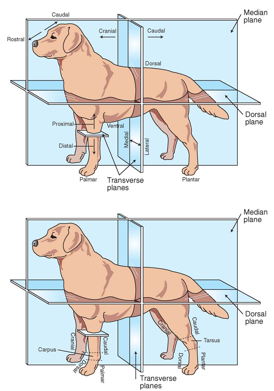
FIGURE 1-1 Anatomic pl anesof reference and Jirmctionalterms. (Modified SromMcBride DF: teaming veterinary terminology, ad 2, St Louic, 2002, Mocby.)
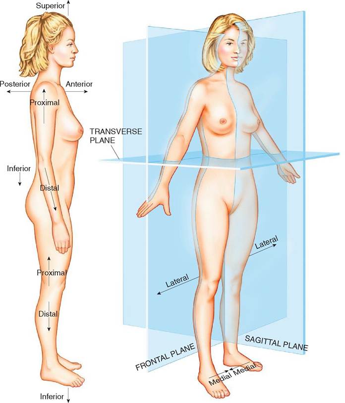
FIGURE 1-2 Directions and planes of the human body. (From Thibodeau GA, Patton KT: Anatomy and physiology, ed 8, St Louis, 2013, Mosby.)