Anatomical nomenclature
As with any field of science, anatomy has its own language. It is necessary to know this language to describe structures and events in a precise and accurate manner. When trying to describe the location of the femur, simply saying that it is "in the back leg and located before the tibia and fibula" will not suffice.
Directional and positional terms
Anatomical terms are used to describe an animal that is in its normal anatomical position. In the case of humans, who are biped (i.e., walk on two legs), this means standing with the arms hanging by the side and the palms rotated forward. For animals that are quadruped (i.e., walk on four legs), anatomical position entails standing on all four limbs.
Positional and directional terms are presented in Table 1.1. The use of such terms allows for more precision while using fewer words to describe body structures. For example, one might say, "The knee is located on the front leg approximately halfway between the trunk and the hoof." With directional and positional terms, one can say, "The knee is located distal to the humerus and proximal to the radius and ulna, in the middle of the front leg."
These terms can have different meanings when referring to humans as opposed to animals. While dorsal and posterior mean toward the back or spinal column in humans, dorsal means toward the spinal cord in a quadruped, while posterior means toward the tail.
Body planes
When talking about anatomical locations, it is necessary to take into account the three-dimensional nature of an animal. The body can be sectioned, or cut, in all three planes. Knowing which plane one is observing when looking at a cross section gives knowledge of the location of various structures. Looking at anatomical planes has become common in the many television crime and medical mystery shows that show images from various magnetic resonance imaging (MRI) scans.
Using the horse as an example, the terms are further depicted in Figure 1.7.A sagittal plane divides the body into right and left parts along the longitudinal axis (Table 1.2; Fig. 1.7). If the plane is exactly along the midline of the longitudinal axis, it is said to be a median, or midsagittal, plane. Any sagittal plane other than the midsagittal is said to be a parasagittal (para = near) plane.
A frontal (dorsal) plane runs longitudinally and passes through the body parallel to its dorsal surface and at a right angle to the median plane. In other words, it divides an animal into a dorsal and ventral portion and runs parallel to the ground. In humans, such a plane runs perpendicular to the ground.
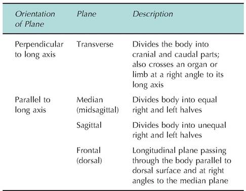
Table 1.2. Body planes.
| Table 1.1. Directional and positional terms. | ||
| Term | Meaning | Example |
| Dorsal | Toward the back; also, below the proximal ends of the carpus and tarsus, dorsal means toward the head (i.e., dorsal replaces cranial) | The vertebral column is dorsal to the sternum. |
| Ventral | Toward the belly | The udder is ventral to the tail. |
| Cranial | Toward the head | The neck is cranial to the tail. |
| Caudal | Toward the tail | The tail is caudal to the head. |
| Rostral | Part of the head closer to the nose | The beak is rostral to the ear. |
| Proximal | Near the trunk or origin of the limb | The elbow is proximal to the ankle. |
| Distal | Farther from the trunk | The ankle is distal to the elbow. |
| Palmar | Below the proximal ends of the carpus, palmar replaces caudal | The dewclaws are on the palmar surface of the forelimb. |
| Plantar | Below the proximal ends of the tarsus, planar replaces caudal | The dewclaws of the hind limb are on the plantar surface of the foot. |
| Medial | Toward the longitudinal axis (midline) | The sternum is medial to the limbs. |
| Lateral | Away from the longitudinal axis | The scapula lies lateral to the spine. |
| Superficial | Nearer the body surface | The skin is superficial to the ribs. |
| Deep | Farther from the body surface | The heart is deep to the ribs. |
| Axial and abaxial | Restricted to the digits, these terms indicate position relative to the longitudinal axis of the limb; axial and abaxial are closer and further to the longitudinal axis, respectively | The lateral edge of the hoof is abaxial to the phalanges. |
Chapter 1
A transverse plane runs perpendicular to the long axis of the structure. A transverse plane can divide an animal into a cranial and caudal half, or it can divide a limb into a proximal and distal section.
Body cavities and membranes
A median view of an animal will reveal two cavities, the dorsal and ventral. The dorsal cavity protects the brain and spinal cord and contains the cranial cavity within the skull, and the vertebral, or spinal, cavity that is found within the vertebral column. The brain and the spinal cord are continuous; therefore, the cranial and vertebral cavities are also continuous.
When looking down the longitudinal axis, the trunk of the animal can be divided into three cavities. The thoracic cavity is surrounded by the ribs and muscles of the chest. It can be further subdivided into the pleural cavities, each of which houses a lung, and the mediastinum, which is located medially between the lungs and contains the pericardial cavity. The mediastinum also houses the esophagus and trachea.
The abdominopelvic cavity is separated from the thoracic cavity by the diaphragm. The abdominopelvic cavity has two components: the abdominal cavity that contains, among others, the stomach, intestines, spleen, and liver, as well as the more caudal pelvic cavity. The pelvic cavity is surrounded by the bones of the pelvis, and contains the bladder, part of the reproductive organs, and rectum.
The walls of the ventral body cavities, as well as the surface of the visceral organs, are covered by a thin, double-layer membrane called the serosa, or serous membrane. The portion of the serosa lining the body cavity is called the parietal (pαrie = wall) serosa, while the portion lining the organ is the visceral serosa.
The best way to visualize the relationship between the two layers of the serosa is to imagine pushing your
Chapter 1

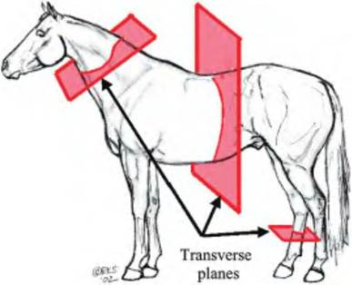
Fig. 1.7. Planes of the body. The three major planes (frontal, transverse, and sagittal) are shown.
fist into an inflated balloon.
The layer of the balloon closest to your fist would be equivalent to the visceral serosa, while that part of the balloon on the outside would represent the parietal serosa. The two serosal membranes each secrete serosal fluid into the space between the two layers. This fluid acts as a lubricant to reduce the friction between the parietal and visceral serosa as they slide across one another. This is important when one considers how often the heart beats or the lungs inflate, during which time the visceral and parietal serosa slide across one another.The serosa membranes have specific names depending on their locations. When found surrounding the heart, it is called the pericardium (peri = around + kardia, heart). Therefore, the parietal pericardium lines the pericardial cavity, while the visceral pericardium adheres to the heart. The pleura adheres to the lungs and lines the thoracic cavity, whereas the peritoneum lines the abdominopelvic cavity and adheres to the visceral organs.
Review questions and answers are available online.
References
Bernard, C. 1965. An Introduction to the Study of Experimental Medicine. Great Books Foundation, INC., Chicago, Illinois.
Cannon, W.B. 1932. The Wisdom of the Body. Norton, New York.