Baylisascaris spp. Larval Migrans
The natural host of Baylisascaris procyonis is the raccoon. However, when an unnatural host such as a rabbit (or human) accidentally ingests infective eggs, a devastating cerebrospinal disorder may result.
Hay or bedding contaminated with raccoon feces containing B. procyonis eggs is the usual source of the parasite. Following passage in raccoon feces, embryonation requires approximately 30 days before the eggs are infective. Eggs will remain infective for at least a year under appropriate environmental conditions. Following the accidental ingestion of embryonated eggs, the larvae are released in the intestine and undergo aggressive somatic and pulmonary migration. Larvae have a tropism for the brain stem. Typical neurological signs include torticollis, ataxia, circling, opisthotonus, and recumbency. If not euthanized, animals usually die as a result of the unremitting nervous signs. In addition, B. columnaris, the ascarid of skunks, may also cause similar disease, but is less common.Pathology
Multiple, circumscribed, raised white nodules up to 1.5 mm in diameter may be found in the subepicardial and subendocardial regions of the heart and the serosal surface of the liver. Microscopic examination of the visceral lesions reveals focal granulomas, with mononuclear cells and heterophils infiltrating the area. Remnants of the parasite are often present within these lesions. In the central nervous system, lesions are most often present in the gray and white matter in the brain stem and cerebellar regions, but the cerebrum, including the hippocampus, may be involved. Sites of parasitic migration are characterized by extensive mal- acia and astrogliosis. Large numbers of Gitter cells and gemistocytic astrocytes may be present in lesions interpreted to be of several days' duration. Infiltrating inflammatory cells include lymphocytes, macrophages, eosinophils, and heterophils.
Within the neuropil adjacent to the lesions, nematode larvae can be identified with characteristic excretory columns and lateral alae (Fig. 6.69). Because of active migratory behavior, larvae may not be found in association with inflammation, but can be found in other regions that have yet to develop an inflammatory response. Thus, if larval migrans is suspected, multiple tissue sections may need to be examined. The primary differential diagnosis is Encephalito- zoon cuniculi infection, but that organism tends not to target the brain stem, and can be identified with tissue Gram stains.Capillaria hepatica Infestation
Capillaria hepatica infects many species, including wild lagomorphs. It was observed in the livers of laboratory rabbits purchased from a commercial supplier in the United Kingdom. Gross findings included irregular white or yellow patches, streaks or small nodules visible
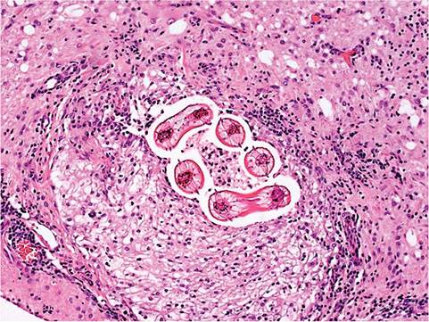
FIG. 6.69. Cerebral Baylisascaris infestation in a New Zealand White rabbit. Note the focus of malacia and inflammation containing multiple cross sections of ascarid larvae with characteristic lateral alae.
on the surface, and cut sections of liver. Lesions included portal inflammation, dilated bile ducts, and fibrosis. The hepatic parenchyma contained multiple granulomas infiltrated with macrophages, eosinophils, and lymphocytes in association with multiple double-operculated ova. In 1 report, ova were also present in the bile ducts and gall bladder of infected rabbits. Wild rodents are the most common definitive hosts for C. hepatica.