BEHAVIORAL, PHYSIOLOGIC, AND ANATOMIC FEATURES
Behavioral Features
Adult male guinea pigs are called “boars,” females “sows,” and infants “pups.” Guinea pigs live in harems with a strong male dominance hierarchy and a loose female hierarchy.
They tend to live in family units centered around an alpha male. Mature boars, particularly if strangers, will fight savagely, sometimes with fatal outcome. Sows will also fight on occasion. Guinea pig activity tends to be crepuscular. Vocalization is well developed and complex. They respond to sudden auditory stimulation and unfamiliar surroundings by freezing in place (immobility response), whereas sudden movement will elicit a random stampede (scatter response), which may result in injury to young animals. Guinea pigs eat frequently and unlike most rodents, do not cache their food or burrow, although wild cavies will make use of borrowed burrows. They require a constant source of water and tend to contaminate their water sources with ingesta as they drink. They do not lick sipper tubes without training, a consideration that can lead to dehydration and death. They are indiscriminate defecators and renowned for their tendency to sit in and soil their food bowls. Guinea pigs are polyestrus, with breeding activity occurring year-round. Sows do not make nests. The gestation period is 59-72 days, depending upon litter size, which generally ranges between 1 and 6 pups. Sows have a postpartum estrus within 2-10 hours after farrowing. Pups are highly precocious at birth and are fully furred and mobile, with eyes completely open. At parturition, both boars and sows assist in grooming pups and eating placentas, and lactating sows will nurse unrelated pups. Newborn pups do not receive much maternal attention, other than anogenital grooming, which stimulates defecation and urination. Cannibalism or consumption of aborted fetuses and stillborn pups does not occur. Pups are normally weaned within 3 weeks but can be weaned as early as 3-4 days if anogenital stimulation is provided. Nervous in temperament, guinea pigs may refuse to eat or drink for some time following any significant change in location, feed, or management practices.Pathology of Laboratory Rodents and Rabbits, Fourth Edition. Stephen W. Barthold, Stephen M. Griffey, and Dean H. Percy. © 2016 John Wiley & Sons, Inc. Published 2016 by John Wiley & Sons, Inc.
Unique Physiologic Features
Unlike other rodents and similar to primates, guinea pigs require dietary sources of vitamin C due to deficiency in l- gulonolactone oxidase. Commercial diets that are formulated for guinea pigs are supplemented with vitamin C, but attention must be paid to the expiration date and proper storage due to instability of this vitamin. Provision of leafy green vegetables is often used to supplement guinea pig diets, which is thoroughly enjoyed, but poses a risk for introduction of pathogens to guinea pig populations. Similar to rabbits, guinea pigs absorb calcium through their intestine in proportion to the amount in the diet, and depend upon renal excretion for calcium balance (see Rabbit Chapter 6, “Physiologic Features”).
Anatomic Features
External Features
Guinea pigs have 4 toes on their front feet and 3 toes on their hind feet with hairless footpads. There is a region behind the ears that normally lacks hair that can be misconstrued as alopecia. They possess a vestigial tail with a supracaudal scent gland (coccygeal gland or “grease gland”). In addition, guinea pigs have apairof perineal scent glands (anal or perianal glands) and sebaceous glandular tissue in the penile sheath and skin of the perineal sacs. The perineal sacs, which are larger in boars, consist of bilateral skin diverticula that surround the anogenital region and secrete and accumulate a creamy substance for scent marking. Dermal sebaceous glands are abundant along the dorsum and perineal regions, particularly in boars.
Old or obese guinea pigs that are incapable of suitable grooming may take on a greasy appearance. Guinea pigs have a single pair of inguinal mammary nipples, but supernumerary nipples are frequent. The external genitalia are unique, and pose a challenge for the inexperienced when attempting to sex young pups. Sex identification in pups is facilitated by gentle pressure above the prepuce, which causes partial extrusion of the penis in males. The inguinal canals are open and the scrotum of boars is represented as scrotal pouches lateral to the prepuce and anus. The urethral opening of sows is outside the vagina, and the vaginal orifice is closed by a vaginal closure membrane during anestrus and pregnancy (see “Reproductive System”).Lymphoid and Hematopoeitic Systems
The counterpart of the neutrophil in guinea pigs is the heterophil or pseudoeosinophil, due to distinct eosinophilic cytoplasmic granules. Lymphocytes are the
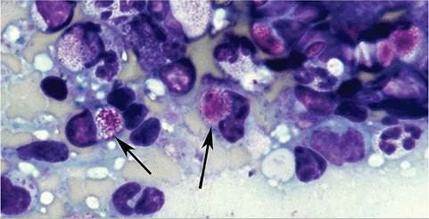
FIG. 5.1. Impression smear of spleen from an adult female guinea pig, illustrating Kurloff cells (arrows). Note the large, finely granular Kurloff bodies within the cytoplasm of these mononuclear cells.
predominant leukocyte in the peripheral blood, and both small and large forms are normally found. Up to 4% of circulating white blood cells may be Kurloff (Foa- Kurloff) cells. Kurloff cells are unique innate mononuclear leukocytes with natural killer (NK) cell activity that are found routinely in certain tissues in guinea pigs and have also been documented in capybaras. The cells contain a finely fibrillar to granular structure (Kurloff body) 1-8 μm in diameter within a cytoplasmic vacuole, with displacement of the nucleus (Fig. 5.1). The intra- cytoplasmic material is PAS positive and stains positive for fibrinoid material with the Lendrum stain. On ultra- structural examination, inclusions are membrane bound, and cytoplasmic organelles in these cells are consistent with secretory activity.
In nonpregnant animals, Kurloff cells are located primarily in the sinusoids of the spleen, particularly in sows, and in stromal tissues of the bone marrow and thymus. Large numbers of Kurloff cells accumulate in pulmonary capillaries (Fig. 5.2). They are not normally found in lymph nodes.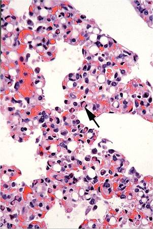
FIG. 5.2. Kurloff cells (arrow) within pulmonary capillaries of an adult female guinea pig.
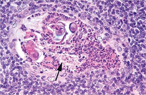
FIG. 5.3. Thymus from a young guinea pig. Note the prominent Hassall's corpuscle (arrow) containing desquamated epithelium, cellular debris, and heterophils, which is a normal feature of the thymus in this species.
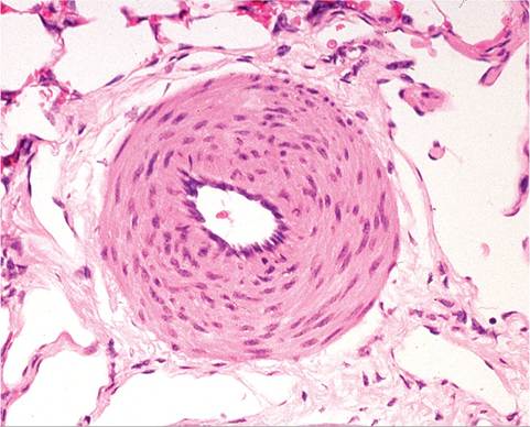
FIG. 5.4. Pulmonary artery in the lung of a normal guinea pig, illustrating the thick smooth muscle of the tunica media.
Kurloff cells are rarely seen in newborn guinea pigs, but are present in relatively large numbers in adult sows. Their numbers fluctuate with the stage of the estrous cycle. Increased numbers are usually present in the peripheral blood during pregnancy. In addition, large numbers of these cells aggregate in the placental labyrinth in pregnant sows. Kurloff cells have been shown to release material into the trophoblast and fetal endothelium of the placental labyrinth. In vitro studies have demonstrated that this material has a toxic effect on macrophages. It has been suggested that Kurloff cells may play a role in preventing maternal rejection of the fetal placenta during pregnancy.
Thymic tissue is present in the cervical region, and accessory thymic tissue is frequently associated with the parathyroid glands. Hassall's corpuscles are very prominent in guinea pigs, with exfoliation of squamous cells and infiltration of heterophils. Degenerate thymocytes are frequently observed in close association with Hassall's corpuscles (Fig.
5.3). These areas of degeneration may evolve into small cysts. The thymus involutes with age, which is essentially complete by 1 year. Maternal antibody is transferred in utero during late gestation through the yolk sac splanchnopleure.Respiratory System
Guinea pigs, like other rodents and rabbits, are obligate nasal breathers, so that obstruction of nasal passages with exudate may present as dyspnea. Pulmonary arteries and arterioles have well-developed smooth muscular thickening of the tunica media (Fig. 5.4), which can be misconstrued as an abnormal finding. Smooth muscle is also prominent around pulmonary veins. Furthermore, longitudinal orientation of pulmonary arteries reveals that smooth muscle is arranged in unique segmental bulges, reminiscent of sphincters (Fig. 5.5). Thus, depending upon level of section, arteries may either appear to be surrounded by a lot of smooth muscle or none at all. Unlike other rodents, cardiac muscle does not surround pulmonary veins within the lung. Larger airways are surrounded by prominent concentric bands of smooth muscle. The contraction of the peribronchial muscle may result in marked distortion, thickening, and sloughing of the respiratory epithelium that lines affected airways. Such artifacts have been interpreted to be neoplastic by the untrained observer. Clara cells are the prevalent cell type lining bronchioles, but they are absent in the trachea and larger bronchi.
Aggregations of lymphocytes in the adventitia of pulmonary vessels are a common incidental finding in guinea pigs. Microscopic foci have been observed in animals as young as 5 days of age, but nodular aggregates are more common in older animals. These perivascular changes are normally found only in the lung. At necropsy, close examination may reveal circumscribed, pale, pinpoint
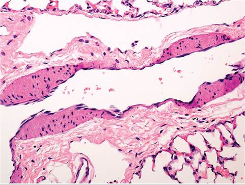
FIG. 5.5. Longitudinal section of a pulmonary artery in a normal guinea pig, revealing the unique segmental bulges of smooth muscle, which are reminiscent of sphincters.
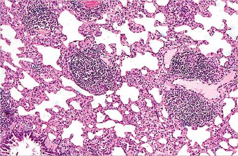
FIG. 5.6. Lung from an adult guinea pig with prominent perivascular lymphoid aggregates. These infiltrates are commonly present in the adventitia of small pulmonary vessels of adult guinea pigs.
subpleural foci up to 0.5 mm in diameter. On microscopic examination, concentric to eccentric aggregations of small- to medium-size lymphocytes are oriented around small arteries and veins (Fig. 5.6). Nodules are focal to segmental in distribution in the perivascular regions. There may be focal to diffuse infiltrates of lymphocytes and thickening of the alveolar septa in some animals, but the airways and alveoli are free of exudate in the typical cases. Ultrastructural studies have revealed morphologically normal lymphocytes, and there was no evidence of viral agents associated with the cellular infiltrates. The lymphoid nodules have been ascribed to a variety of antigenic stimuli, but the pathogenesis and significance of these changes are not well understood. If the lesions are accompanied by adjacent alveolar inflammation, they are likely to be associated with a disease process.
Bony spicules (osseous metaplasia) have been observed in the lung in guinea pigs. Similar changes have been seen in other species, such as the rat and hamster. They are composed of dense, lamellar bone, with varying degrees of mineralization. There is usually no or minimal reaction in the adjacent alveolar septa. They have been interpreted as inhaled fragments of bone of dietary origin, but it is more likely that they are foci of osseous metaplasia. Large numbers of metaplastic osseous foci, including well-differentiated bone marrow, have been observed in the lungs of guinea pigs following X-irradiation.
Gastrointestinal System
Guinea pigs are monophydont with open-rooted, continuously growing (elodont) incisors and cheek teeth. Their dental formula is I1/1, C0/0, P1/1, M3/3. Cheek teeth grow slightly inward, predisposing them to interfere with mastication if overgrown. Guinea pigs have a simple stomach without a nonglandular portion. They are hindgut fermenters with a large cecum that holds approximately 65% of the enteric digesta. The cecum has 3 linear taeniae and taeniae coli run the entire length of the colon. Unlike most other species, the guinea pig cecum is located on the left side of the abdominal cavity.
In contrast to the rabbit, the only macroscopic gut- associated lymphoid tissues are Peyer's patches. Guinea pigs engage in cecotrophy, with production of mucoid vitamin-rich cecotropes, but they do not have the more complex retropulsive system of rabbits. Guinea pigs digest 34% of crude fiber compared to only 10% by rabbits fed the same diet.
Urinary System
As with rabbits, renal excretion of calcium is the major means of calcium homeostasis in guinea pigs. Guinea pig urine, like that of rabbits, is therefore normally thick and cloudy with numerous crystals visible on microscopic examination. This material may accumulate as “sludge” in the bladder, but obstruction due to sludge is not common, whereas calculi may develop and result in obstructive uropathy. Guinea pigs enjoy alfalfa, but its high calcium content may predispose them to urolithiasis.
Reproductive System
Boars, like other rodents, are endowed with a number of accessory sex glands, including large seminal vesicles, coagulating glands, prostate, and bulbourethral glands. They produce a copulatory plug upon ejaculation. The glans penis has numerous cornified scales or spurs. The ventral aspect of the glans has an intromittent sac with additional scales and 2 horny styles that evert upon erection. Sows have a vaginal closure membrane that is unique to hystricomorph rodents (Fig. 5.7). During pregnancy and anestrus, the vaginal orifice is sealed with a membrane covered with stratified squamous epithelium on the internal and external surfaces. The
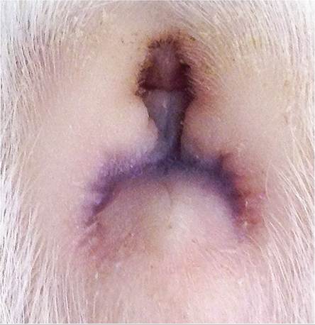
FIG. 5.7. Vaginal closure membrane in an anestrus guinea pig sow. During anestrus and pregnancy, the vaginal orifice is sealed with an epithelial membrane, which is unique to hystricomorph rodents. (Courtesy M. Hunrath)
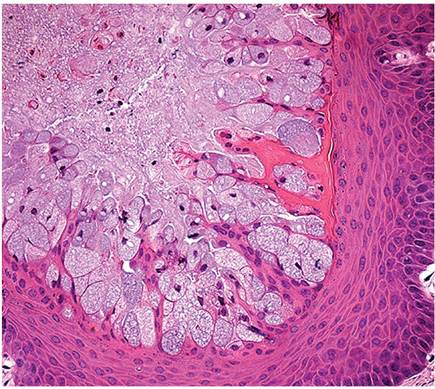
FIG. 5.8. Vaginal mucosa of a normal guinea pig sow, illustrating mucinous metaplasia of stratified squamous epithelium.
membrane ruptures when the vulva swells prior to parturition and during estrus. During pregnancy, the vaginal lumen fills with mucoid fluid produced by mucoid metaplasia (mucification) of the cornified epithelium (Fig. 5.8). This occurs in other rodents, but is particularly pronounced in guinea pigs. The guinea pig uterus is bicornuate, with a very short uterine body and a single cervical os. Placentation is discoidal and hemomonocho- rial. Its structure has favored its study as a model of human placentation. Placentation includes a subplacenta, which is unique to hystricomorph rodents. It is an extension of the chorion into the floor of the central placenta on the maternal side of the main placenta and is separated from the main placenta by a band of fetal mesenchyme.
Musculoskeletal System
The pubic symphysis generally remains fibrocartilaginous throughout life, but may completely ossify in older boars. Preparturient sows elaborate the peptide hormone relaxin, which is produced by the corpus luteum and placenta. This allows relaxation of the pubic ligament, which features leukocytic infiltration, degradation of collagen, and angiogenesis, resulting in increased weight and length of the ligament (Fig. 5.9). This allows passage of the exceptionally large and precocious fetuses. This process is less efficient in older sows, whose pubic symphyses tend to become partially ossified.