Bones and skeleton
Markings on bones
The surface of bone is seldom smooth. Instead, various depressions, bumps, and ridges serve as sites where muscles and tendons originate or attach and blood
vessels and nerves travel.
These various markings are shown in Table 6.1 and Figure 6.12. Learning the terms is helpful when studying the origins and insertions of muscles.Skeleton
The skeleton includes all the bones of the body. These bones and their articulations have been altered during evolution to accommodate various functions. Therefore, the skeleton is an excellent example of the complementary nature of form and function. The skeletons of various species are shown in Figure 6.13. Most of the remainder of the chapter is concerned with mammals; however, a brief discussion of features unique to avian species will be included at the end of the section on the skeleton.
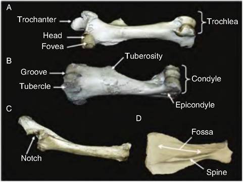
Fig. 6.12. Marking on bones. Various bovine bones are shown, including the femur (A), humerus (B), radius-ulna (C), and scapula (D), which are labeled to illustrate bone markings.
Table 6.1. Bone markings.
| Term | Description | Example |
| Projections, Depressions and Openings Where Muscles and Ligaments Attach | ||
| Crest | Narrow ridge of bone; usually prominent | Iliac crest |
| Epicondyle | Raised area on or above a condyle | Lateral epicondyle of the humerus |
| Fossa | Shallow depression, often serving as an articular surface | Olecranon and radial fossae of the humerus |
| Line | Narrow ridge of bone; less prominent than a crest | Gluteal line on wing of ilium |
| Process | Generally any bony prominence; sometimes used to name specific prominences | Crest, spine, trochanter, tubercle, tuberosity, etc.; olecranon process |
| Ramus | Armlike bar of bone | Ramus of the mandible |
| Spine | Sharp, slender, often pointed projection | Spine of the scapula |
| Tuberosity | Large rounded projection | Deltoid tuberosity of the humerus |
| Trochanter | Very large, blunt, irregular-shaped process; found only on the femur | Trochanter of the femur |
| Tubercle | Small rounded projection or process | Greater tubercle of the humerus |
Projections That Help Form Joints
| Condyle | Rounded articular projection | Occipital condyle of the skull |
| Cotyloid | A deep articular depression | Acetabulum of the hip joint |
| Facet | Smooth, nearly flat articular surface | Superior costal facet of the vertebrae |
| Head | Bony expansion carried on a narrow neck | Head of the femur |
| Trochlea | A pulley shaped, articular structure | Trochlea of the femur |
Depressions and Openings Allowing Blood Vessels and Nerves to Pass
| Fissure | Narrow, slit-like opening | Palatine fissure |
| Foramen | Round or oval opening through a bone | Foramen magnum |
| Fovea | A shallow, Iionarticular depression | Fovea capitis on the head of the femur |
| Incisure | A notch-shaped depression at the edge of a bone | Semilunar notch of the ulna |
| Meatus | Canal-Iike passageway | External auditory meatus |
| Sinus | Cavity within a bone, filled with air and lined with mucous membrane | Nasal sinuses |
| Sulcus | Furrow-Iike groove | Brachial groove of the humerus |

Fig. 6.13.
Skeletons. (A) Cow. (B) Horse. (C) Pig. (D) Dog.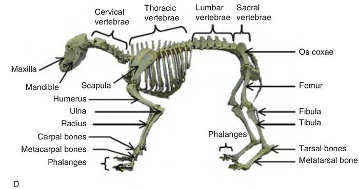
Fig. 6.13. Continued
Functions of the skeletal system
The skeleton has five primary functions:
1. Support. The skeletal system provides the structure to which the bones attach, as well as the structural support for the entire body.
2. Storage of minerals and lipids. The bones provide major storage for various minerals, particularly calcium. In addition, the bones contain a substantial amount of lipid.
3. Blood cell production. The bone marrow is a site of formation for all types of blood cells.
4. Protection. The vital organs of the body are protected by the skeletal system. The ribs surround the visceral organs, whereas the central nervous system is encased within the skull and spinal cord.
5. Leverage. Many of the joints of the body act as levers, therefore assisting with movement.
Skeletal cartilage
Types of cartilage
The skeleton begins as cartilage and fibrous membranes, but then is replaced with ossified tissue as the animal develops through gestation. Cartilage contains no nerves or blood vessels and is surrounded by a layer of dense irregular connective tissue called the perichondrium. Blood vessels found within the perichondrium provide nutrients for the chondrocytes within the cartilage.
There are three types of cartilage found in the skeleton. Hyaline cartilage is the most abundant and provides support and flexibility for the skeleton. The matrix contains only fine collagen fibers. Hyaline cartilage is found (1) on articular surfaces, (2) within costal cartilage connecting the ribs to the sternum, (3) in the respiratory cartilages forming the skeleton of the larynx and reinforcing passageways of the respiratory system, and (4) in nasal cartilages supporting the external nose.
Elastic cartilage contains more elastic fibers than hyaline cartilage. It is therefore better able to withstand bending.
It is found in only two places in the skeleton: (1) the external ear and (2) the epiglottis, which is the flap of tissue that covers the opening of the larynx during swallowing.Fibrocartilage is highly compressible, possessing great tensile strength. It contains approximately parallel rows of chondrocytes with intervening thick collagen fibers. It is found in the menisci within the knee and intervertebral discs.
Growth of cartilage
Cartilage can continue to grow by two processes. Appositional growth occurs when new cartilage forms on the surface of preexisting cartilage. Interstitial growth occurs from inside of the cartilage mass in which lacunae-bound chondrocytes inside the cartilage divide and secrete new matrix, thereby expanding the cartilage from within.
Skeleton classification
The skeleton is divided into the appendicular skeleton and the axial skeleton (Fig. 6.14). The axial skeleton includes the skull, hyoid apparatus, vertebral column, ribs, and sternum. The appendicular skeleton includes the bones of the limbs and limb girdles. The thoracic limb or pectoral limb includes the scapula, humerus, radius, ulna, carpal bones, metacarpal bones, phalanges, and their sesamoid bones. The thoracic girdle or shoulder girdle includes the two scapulae and the clavicle in man, which holds the shoulder laterally, but which is only vestigial in domestic animals.
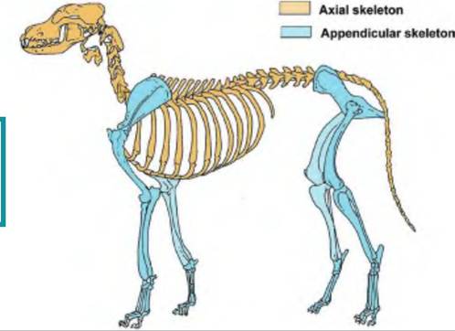
Fig. 6.14. Axial and appendicular skeleton. As shown with this dog skeleton, the axial skeleton includes the bones and cartilage protecting the soft structures in the head, neck, and trunk, and consists of the skull, hyoid apparatus, vertebral column, and thorax. The appendicular skeleton includes the limbs and bones connecting the limbs to the axial skeleton.