Axial skeleton
The skull
The skull is a very complex structure made mostly of flat bones. Except for the mandible that is attached via a movable joint, the bones of the skull are connected by interlocking joints called sutures.
The suture joints are characterized by a saw-toothed or serrated appearance that keeps the bones attached, but allows the cranium to expand and contract while remaining intact.Suture lines are visible between the bones of the skull (Fig. 6.15). The internasal suture is between the two nasal bones while the frontonasal suture separates the frontal bones from the nasal bones. The frontoparietal suture separates the frontal bones from the parietal bones. The nasomaxillary suture separates the nasal bones from the maxillary bones.
The skull contains both cranial and facial bones (Table 6.2). The cranium includes those bones that surround the brain. The cranium consists of the cranial vault, also called the calvaria, forming the superior, lateral, and posterior aspects of the skull, and the cranial base or floor that forms the inferior aspect of the cranium. The cranial base is divided by bony ridges into three distinct fossae: the anterior, middle, and posterior cranial fossa. The cranial bones form the
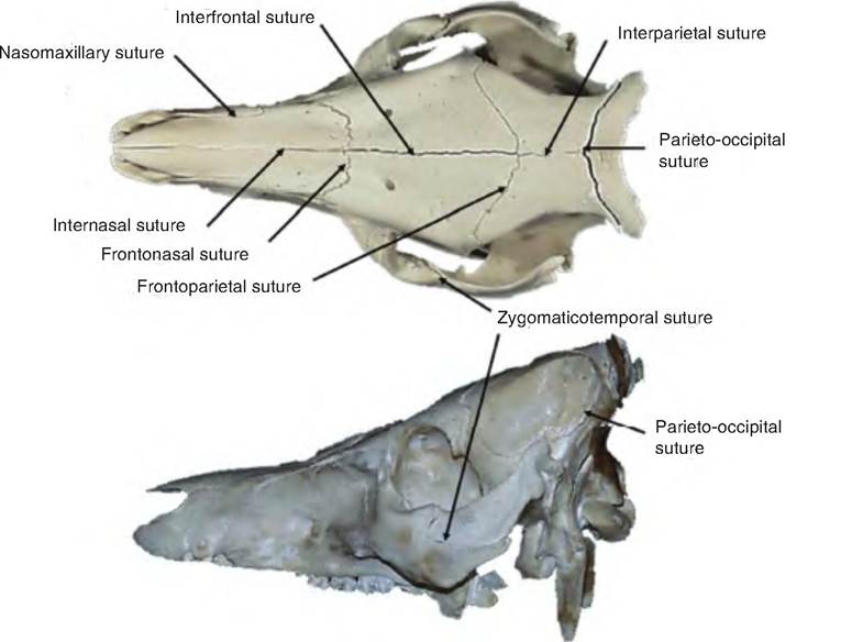
Fig. 6.15. Suture lines of the skull. These lines are shown on top and side view of skull of a pig.
Table 6.2. Bones of the skull.
| Term | Description |
Cranial Bones (Number)
| Frontal (2) | The rostral portion of the roof of the cranial cavity in most domestic species; in the ox, it forms entire roof of cranial cavity |
| Parietal (2) | Along with frontal, forms the roof of the cranial cavity in most domestic animals except ox. |
| Occipital (1) | Forms caudal aspect of the cranial cavity, as well as the skull |
| Temporal (2) | Forms Caudolateral wall of the cranial cavity |
| Sphenoid (1) | Unpaired bone forming floor of cranial cavity; it has several parts, including the body, greater wings, lesser wings, and pterygoid processes |
| Ethmoid (1) | Unpaired bone forming rostral wall of cranial cavity; forms part of the nasal septum, caudal wall of nasal cavity, and part of medial wall of the orbit |
Facial Bones (Number)
| Mandible (1) | The lower jaw |
| Maxilla (2) | Form the upper jaw, and parts of the hard palate, orbits, and nasal cavity |
| Zygomatic (2) | Cranial portion of zygomatic arch; forms part of cheek and orbit |
| Nasal (2) | Along with cranial portion of frontal bone, forms osseous roof of nasal cavity |
| Eacrimal (2) | Forms medial surface of orbit |
| Palatine (2) | Forms part of hard palate along with maxillary and incisive bones |
| Vomer (1) | Unpaired bone forming part of osseous nasal septum |
| Ventral nasal concha (2) | A fragile scroll of bone that increases nasal surface area |
| Pterygoid (2) | Small bones in caudal part of nasopharynx |
| Incisive or premaxillary (1) | Holds upper incisors |
cranial cavity that houses the brain and also provide the site for attachment of head and neck muscles.
The skull contains approximately 85 named openings, including foramina, canals, fissures, and orbits. These provide passageways for the spinal cord, blood vessels, and the 12 cranial nerves to enter and leave the brain.
Cranium
The roof of the cranium is formed by the paired frontal and parietal bones (Fig. 6.16). The caudal aspect of the skull is formed by the unpaired occipital bone. The floor of the cranium is formed by the unpaired sphenoid bone. Finally, the rostral wall of the cranium is formed by the ethmoid bone.
The facial bones include those bones enclosing the nasal and oral cavities. These bones form the structure of the face; contain cavities for special senses, including sight, taste, and smell; provide openings for air and food; secure teeth; and provide attachment sites for facial muscles. The facial region is divided into the oral, nasal, and orbital regions.
The oral region is formed by parts of the incisive, maxillary, and palatine bones, as well as the mandible surrounding the oral cavity. The nasal region is formed by portions of the nasal, maxillary, palatine, and incisive bones that surround the nasal cavity. The orbital region consists of the bony socket holding the eye and is formed by portions of the frontal, lacrimal, palatine, sphenoid, and zygomatic bones. The zygomatic arch, which forms the ventral wall of the orbit, consists of the zygomatic bone and the zygomatic process of the temporal bones. An exploded view of the equine skull is shown in Figure 6.17.
Species differences
Unique to the horse and cat, the interparietal bone is found between the two parietal bones. In other species, this bone is present in the fetus, but fuses with surrounding bones during gestation. In the ox, the frontal bone forms the entire roof of the cranium, whereas the parietal bones help form the roof in other species.
In the dog, three types of skulls are described based on the proportions of the facial bones and cranial cavity (Fig.
6.18):1. Mesaticephalic. Average conformation (e.g., Siberian husky).
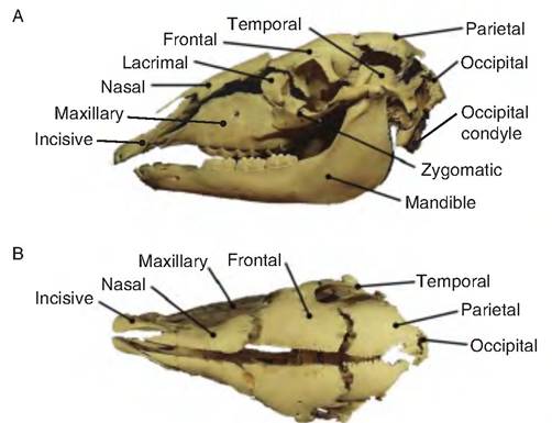
Fig. 6.17. Exploded equine skull. The various bones of the equine skull have been separated, showing side (A) and top (B) view.
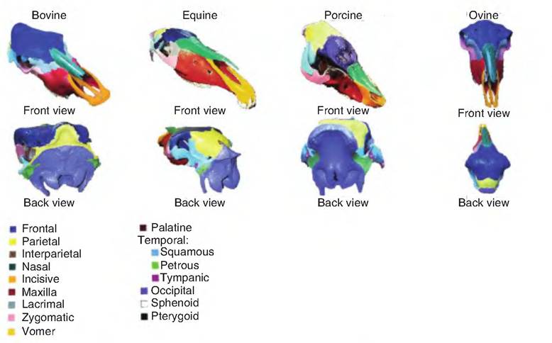
Fig. 6.16. Skulls of various species. The skulls of different species showing main bones.
2. Dolichephalic. Has an elongated facial component (e.g., collie).
3. Brachiocephalic. Has a shorter facial component (e.g., bulldog).
The vertebral column
The vertebral or spinal column, also colloquially (though incorrectly) called the spine (a spine is a pointed projection from any bone), protects the spinal cord, supports the head, and serves as an attachment
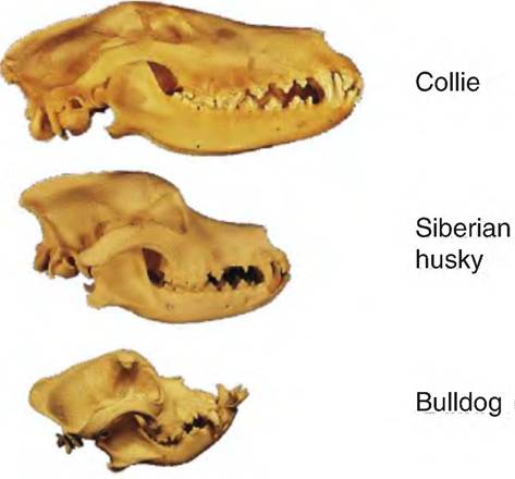
Fig. 6.18. Examples of dog skulls. The three general types of dog skulls are represented. The collie, Siberian husky, and bulldog represent the dolichephalic, mesaticephalic, and brachiocephalic types of skulls, respectively. Note the relatively long facial component on the collie and the short facial component on the bulldog.
site for muscles affecting body movements. The bony column consists of irregular bones connected by joints that are characterized by different degrees of mobility in different regions of the column.
The vertebrae (singular = vertebra) are the irregularly shaped bones making up the vertebral column. Five general regions are described: cervical (neck), thoracic (back), lumbar (loin), sacral, and caudal (tail) vertebrae. Each is named by the first letter of the group followed by the number within the group, for example, Cl, T3, L5, S3, and Ca20. The number of vertebrae typically present in various species is shown in Table 6.3.
Typical vertebrae are shown in Figure 6.19. Features common to all vertebrae include the body, vertebral arch, vertebral foramen, and processes.
The body is the thick, spool-shaped ventral portion of the vertebra. The vertebral body is convex at the cranial end and concave at the caudal end, allowing articulation with the adjacent vertebrae. The vertebral arch is the dorsal portion of the vertebra consisting of two upright pedicles that form the wall of the vertebral foramen. Paired (right and left) half- (or hemi-) laminae project from the pedicles toward the back and, meeting in the midline to complete the lamina, form the roof of the vertebral foramen. In the articulated vertebral column, consecutive vertebral foramen communicate to form the vertebral canal. In the intact state, the spinal cord passes through the vertebral canal.Within each region, the common vertebral features are modified to optimize the ability of each vertebral region to accomplish its function. Regional vertebrae are characterized by features that are more similar to those of others in that same region than to those of the following region. However, the transition from one region to the next is fairly gradual, with the last vertebra of a given region bearing strong resemblance to the first vertebra of the following region.
Potentially, seven processes arise from each vertebra. The dorsal spinous process projects vertically toward the back, and the paired transverse processes
Table 6.3. Number of vertebrae.
| Species | Cervical | Thoracic | Lumbar | Sacral | Caudal |
| Carnivore | C7 | T13 | L7 | S3 | Ca20-24 |
| Pig | C7 | Tl 1 -15 | L6-7 | S4 | Ca20-23 |
| Horse | C7 | T18 | L6 | S5 | Ca15-21 |
| Ox | C7 | T13 | L6 | S5 | Ca 18-20 |
| Sheep | C7 | T13 | L6-7 | S4 | Ca16-18 |
| Chicken | C7 | T7 | L14 (lumbar sacral) |
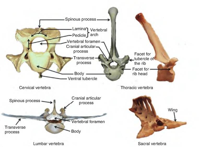
Fig.
6.19. Examples of various vertebrae. Examples of cervical, thoracic, lumbar, and sacral vertebrae are shown. Note that in cervical vertebrae, the spinous process is greatly reduced. Thoracic vertebrae have an exaggerated spinous process, and lumbar vertebrae have an exaggerated transverse process. Sacral vertebrae are generally fused into a single unit, with the processes also tending to fuse into continuous lines rather than individual elevations.extend laterally from each side of the vertebral body. Two pairs (right and left) of cranial and caudal articular processes project from a region-specific area of the lamina to allow adjacent vertebrae to articulate with each other at a second site (the first is at the cranial and caudal articular surfaces of each vertebral body), thus simultaneously allowing greater flexibility and stability than could otherwise be achieved.
The first and second cervical vertebrae are very highly specialized and are unique in form from each other and from any other vertebra. Only these two vertebrae are given specific individual names (Fig. 6.20). The atlas (Cl) supports the head, hence its name (similar to the mythological Atlas, who carried the Earth upon his shoulders). Cranially, the atlas articulates with the occipital condyles of the skull (see Fig 6.17), forming the atlanto-occipital joint. Motion at this joint is a back-and-forth rocking motion, which allows the neck to flex and extend as in nodding "yes." The atlas is unique in that its spinous process is reduced to a bump (the dorsal tubercle), and the body is modified into the ventral arch. The axis (C2) possesses a large ridge-like spinous process. The dens is a heavy, peg-like, cranially directed process that forms a pivoting articulation with the atlas, and allows a twisting motion of the head on the neck, as when shaking the head "no." The dens is developmentally derived from a part of the atlas.
Thoracic vertebrae are generally characterized by robust spinous processes.
The anticlinal vertebra is the one with the most vertically oriented dorsal process. Cranial to this vertebra, the dorsal processes are inclined cranially while those caudal to this vertebra are inclined caudally. The anticlinal vertebra is an important landmark when reading radiographs. The bodies of these vertebrae possess an articular facet at the cranial and caudal end for articulation with the ribs.Lumbar vertebrae are characterized by their massive bodies, shorter spinous processes, and long, flat transverse processes. These vertebrae also lack costal facets since ribs do not articulate with them.
The sacral vertebrae fuse in adult individuals to form the sacrum. Each sacral vertebra has dorsal and ventral foramina allowing the passage of spinal nerves. The wings of the sacrum (Fig. 6.19) articulate with the ilium of the pelvis, forming the sacroiliac joint. This is the single site of connection between the axial skeleton and pelvic limb.
Thorax
The thorax is the bony cavity formed by the sternum, ribs, costal cartilages, and bodies of the thoracic vertebrae (Fig. 6.21). The sternum, or breastbone, is the composite of the unpaired bones (sternebrae) forming the floor of the thorax. Species-specific numbers of sternebrae are as follows: six in pigs, horses, and humans; seven in ruminants, and eight in carnivores. The manubrium is the enlarged first Sternebra, while the xiphoid process is the last Sternebra capped by the xiphoid cartilage. The thoracic inlet is the entrance into the bony thoracic cavity as delineated by the last cervical vertebra, the first pair of ribs, and the sternum.
The ribs consist of long, curved bones that form the lateral wall of the thorax. The ribs can be grouped as follows:
1. Sternal (sometimes called "true") ribs. Articulate directly to the sternum via their costal cartilage. Costal cartilage consists of hyaline cartilage.
2. Asternal (sometimes called "false") ribs. Costal cartilages merge to form the costal arch, which indirectly joins them to the sternum in all domestic species.
3. Floating ribs. End in short costal cartilages that join neither the sternum nor the costal arch, but end "blindly" in the flank musculature. One pair of floating ribs is present in dogs and cats, two pairs in humans and cattle, but none are present in horses.
As shown in Figure 6.22, the proximal part of each rib consists of a head and a tubercle. The head articulates with the caudal and cranial costal fovea of adjacent thoracic vertebrae and the intervertebral disc found in between. The tubercle of the rib articulates with the transverse process of the same numbered vertebra. Between each rib is the intercostal space.