AppendicuIar skeleton
Thoracic limbs
In humans the clavicle braces the shoulder against the sternum, which facilitates climbing and other similar behaviors. In domestic mammals the thoracic limb bears weight and has a much more restricted range of motion, and the clavicle is vestigial.
The proximal end of the thoracic limb begins at the scapula. This "shoulder blade" is a flat, triangular bone in the shoulder region (Fig. 6.23). Together the two scapulae constitute the thoracic girdle.The lateral surface of the scapula presents the spine of the scapula that ends in the acromion, the expanded distal end of the spine of the scapula. The acromion is absent in the horse. The area cranial to the spine is the supraspinous fossa; the area caudal to it is the infraspinous fossa. Most of the medial surface of the scapula is called the subscapular fossa. On the dorsal border of the scapula is the scapular cartilage. On the
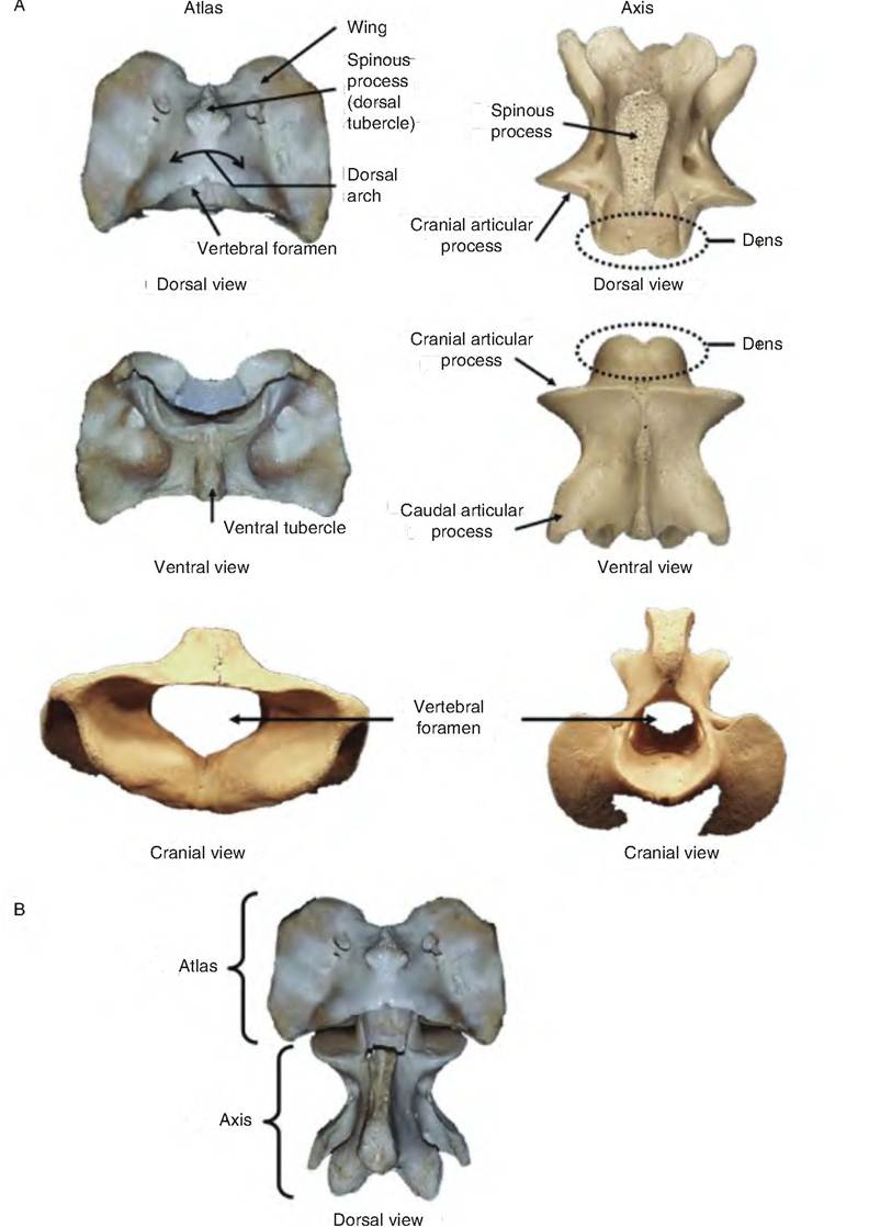
Fig. 6.20. Atlas and axis. The atlas and axis are the first and second cervical vertebrae, respectively. (A) The dorsal, ventral, and cranial views of the atlas and axis of a horse. (B) The articulation between the atlas and axis.
opposite end of the bone, the cavity in which the humerus articulates, is the glenoid cavity. The supraglenoid tubercle, located near the cranial aspect of the glenoid cavity, is the site of proximal attachment (origin) of the biceps brachii muscle. The coracoid process (Greek for "crowlike") is a small process on the medial side of the supraglenoid tubercle where the coracobrachialis muscle arises. Found only in cats, the Suprahamate process is a caudal projection from the acromion.
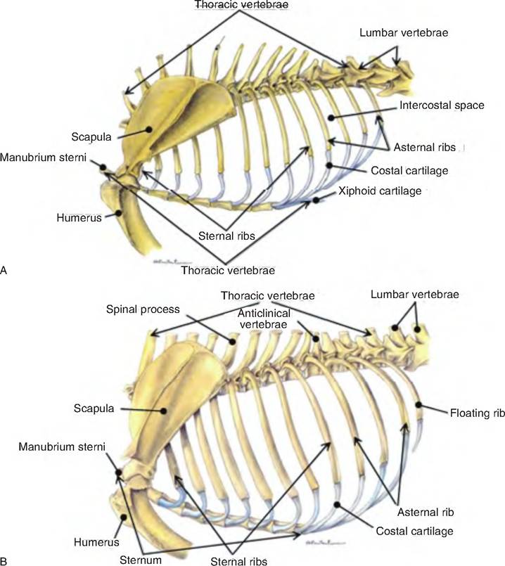
Fig. 6.21. Thorax of different species.
(A) Lateral aspect of cat. (B) Lateral aspect of dog. (Reprinted from Constantinescu, 2002. With permission from the publisher.)The humerus, sometimes called the brachial bone, is the largest bone in the thoracic limb (Fig. 6.24). Proximately, the humerus articulates with the scapula at the glenoid cavity, thereby forming the shoulder joint. Distally, the humerus articulates with the both the radius and ulna Contibuting to the elbow joint.
The head of the humerus is a rounded process articulating with the glenoid cavity. The greater (lateral, major) tubercle is the large process Craniolateral to the head, and can be palpated as the point of the shoulder. The lesser (medial, minor) tubercle is located on the medial side of the head. The bicipital, or intertubercular, groove is a sulcus between the greater and lesser tubercles through which the tendon of the biceps brachii muscle passes. The body of the humerus connects the two epiphyses of the bone. The laterally positioned deltoid tuberosity, to which the deltoid muscles attach, is the largest tuberosity on the bone. The distal end of the bone is called the humeral condyle and includes the humeral capitulum and humeral trochlea that are the two articulating surfaces, two fossae, and the medial and lateral epicondyles. The olecranon fossa is a groove on the caudal surface of the distal end of the humerus in which the olecranon process of the ulna rests. The radial fossa, opposite the olecranon fossa, receives the proximal end of the radius while the elbow is flexed. In the dog, and sometimes the pig, the supratrochlear foramen is a hole that connects the olecranon and radial fossa. Nothing passes through the supratrochlear foramen. In cats only, the supracondylar foramen lies on the lateral surface just proximal to the condyle; the median nerve and the brachial vessels pass through this hole.
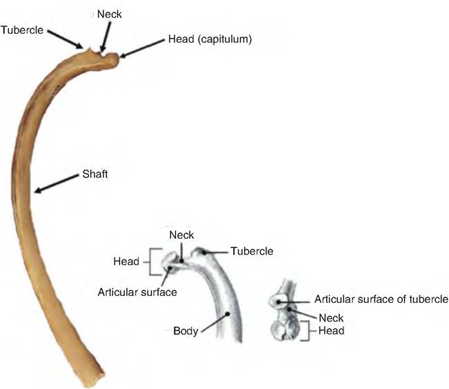
Fig. 6.22.
Typical rib. Typical rib from the mid-thoracic region. (The inserted rib drawing is from Constantinescu and Constantinescu, 2004.)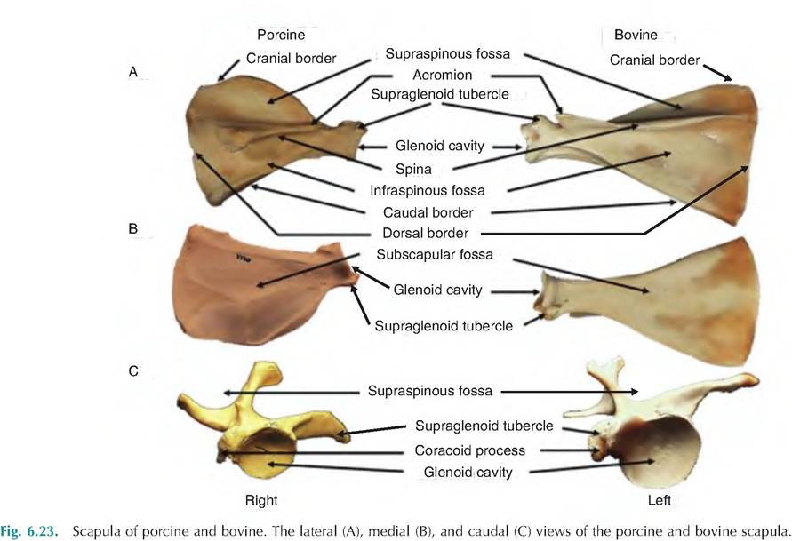
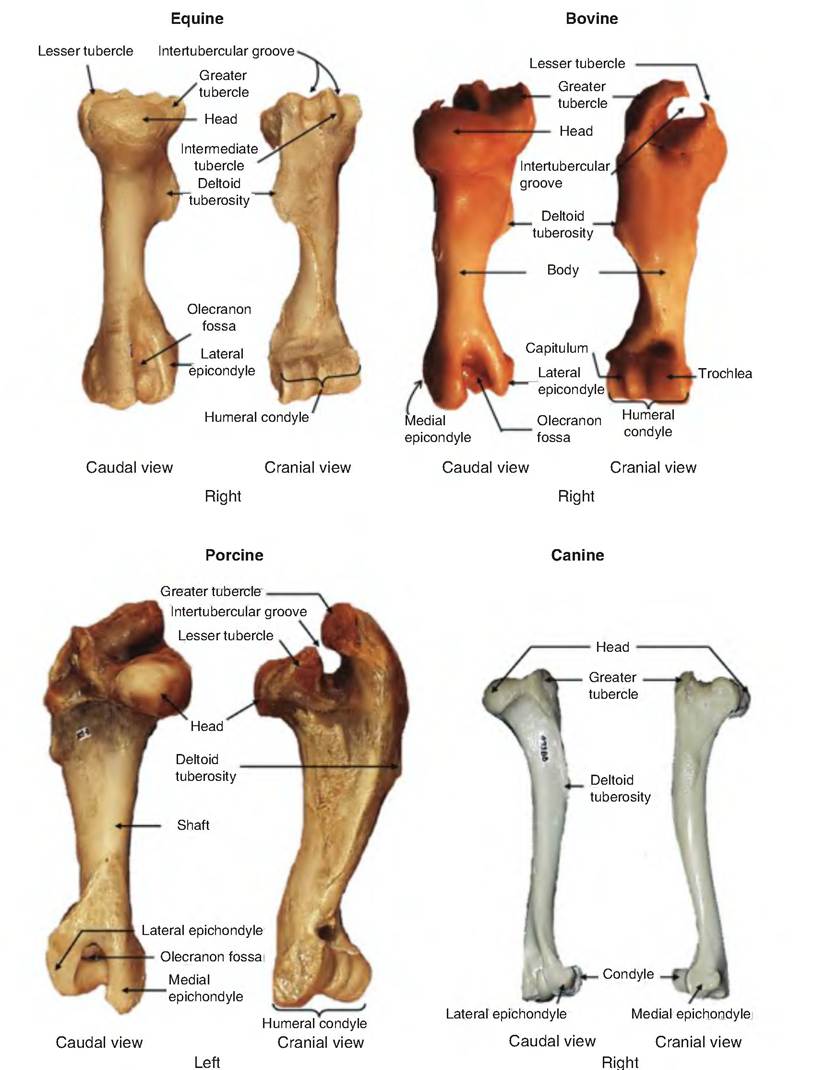
Fig. 6.24. Humerus of various species. The parts of the equine, bovine, porcine, and canine humerus.
The radius is the weight-bearing bone of the forearm (Fig. 6.25). Proximately, the radius articulates with the humerus and ulna at the elbow (the cubital joint); distally, the radial articulation is with the carpal bones and ulna forming the antebrachiocarpal joint. The head of the radius articulates with the capitulum of the humerus, as well as the ulna. The (medial) styloid process lies on the distal end of the radius.
The ulna functions mainly as a site for muscle attachments and formation of the elbow. Proximately, it articulates with the humerus and radius, and distally with the radius and carpal bones. The proximal end of the ulna is called the olecranon process, the point of the elbow where the extensor muscles of the elbow attach. The trochlear notch is the crescentshaped articulation site with the humerus. The distal
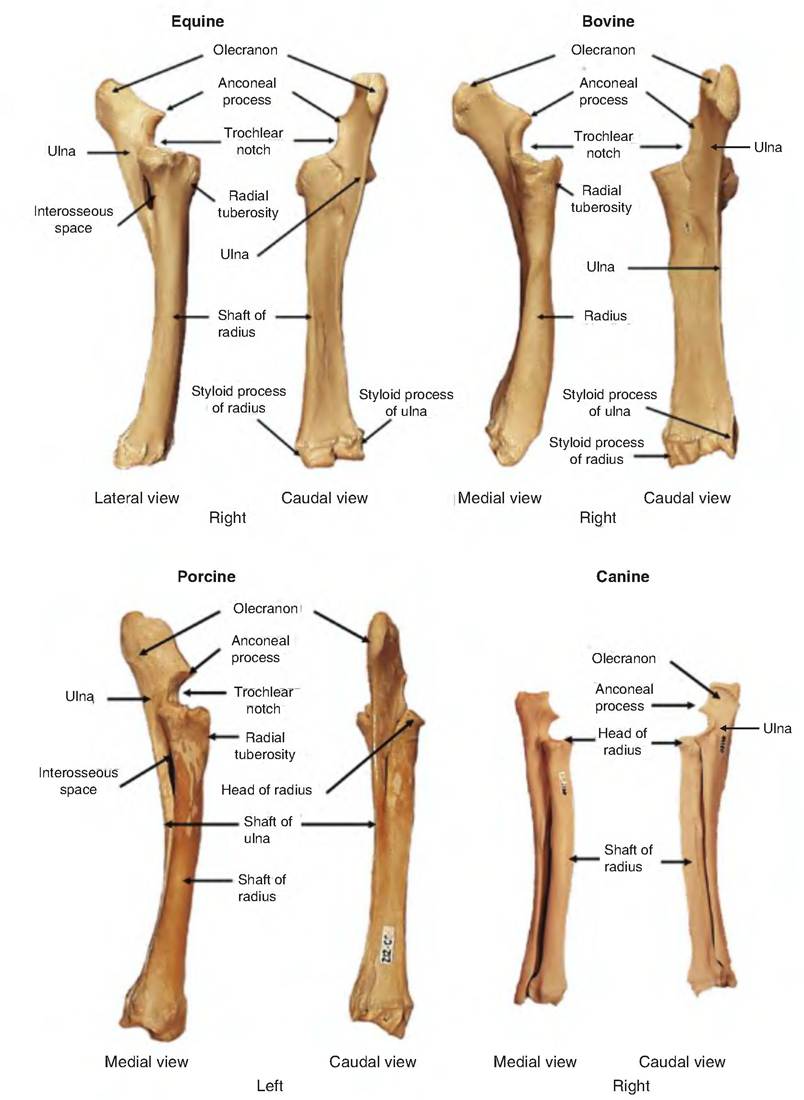
Fig. 6.25. Radius and ulna. The radius and ulna of various species.
end of the ulna also ends in the styloid process, referred to as the lateral styloid process to distinguish it from the radial styloid process.
The radius and ulna fuse in the horse and ruminants. Because they are fused, these animals cannot supinate or pronate (rotate the wrist), and the mannus (hand of humans) is committed to permanent pronation (palm-down position). In contrast, these bones are not fused in carnivores; therefore, these animals can at least partially supinate (turn upward) their paws.
Premature closing of the growth plates in the radius or ulna can cause deviations in these bones resulting in valgus or vargus deviations.
Valgus is a lateral deviation distal to a joint; vargus is a medial deviation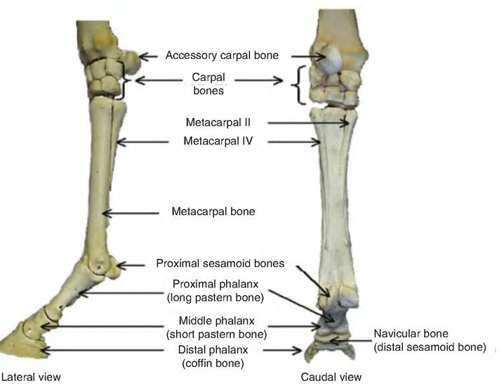
Fig. 6.26. Lower leg, including the mannus, of the horse.
distal to a joint. For example, carpus valgus or carpus vargus are lateral and medial deviations distal to the carpus. Carpal valgus, a lateral deviation of the joints distal to the carpus, is also called "knock-kneed"; carpal vargus, a medial deviation of the bones distal to the carpus, is called "bowlegged."
The distal portion of the thoracic limb is technically the manus (hand), commonly called the forepaw in carnivores (Fig. 6.26). The manus consists of the carpus, metacarpus, and digits. Each digit is a complete toe, including two or three separate bones called sesamoid bones, which are associated with the joints between each metacarpal bones and digit, as well as some of the joints between phalanges (Interphalangeal joints).
The carpus, the wrist of humans, consists of two transverse rows of carpal. The number of carpal bones varies among species. The pig and horse have eight carpal bones, although the first carpal bone in the distal row is often absent in the horse. Dogs and cats have seven carpal bones due to the fusion of two of the carpal bones in the proximal row. Ruminants have six carpal bones since the first carpal bone is absent and the second and third are fused.
The metacarpal (MC) bones are located between the carpus and digits (toes). Potentially five are present, and they are numbered I-V from medial to lateral. Species differ in the number and relative size of metacarpal bones due to absence or fusion of these bones as related to which are weight-bearing. Generally, paired proximal sesamoid bones are present on the palmar (ground-facing) surface at the metacarpophalangeal joint (junction of metacarpal bone and its associated digit). In dogs, only a single sesamoid bone is present for the dewclaw.
Carnivores have five metacarpal bones because all five toes are present. The first metacarpal bone is much shorter than the others because digit I (the "dewclaw") is not weight bearing.
The pig has four metacarpal bones since MC I is absent. The third and fourth metacarpal bones are weight bearing and therefore the longest, while the second and fifth are somewhat shorter and non- weight-bearing on solid ground.
The horse has three metacarpal bones, with MC I and V absent. The second and fourth metacarpal bones are commonly called splint bones because they are greatly reduced in size and end proximal to the digit. The rounded distal ends of each splint bone are called the buttons of the splints. The large, heavy third metacarpal is called the cannon bone.
The digits of the forelimb correspond with the fingers of humans. Like the metacarpal bones (which support the digits), potentially five digits are present and again are numbered from medial to lateral. Their numbers correspond to their supporting metacarpal bones. However, the number of digits present varies by species.
Each digit contains multiple bones called phalanges. The first digit (when present) possesses two pha- Ianges, while the remaining digits possess three. The phalanges are named proximal phalanx, middle phalanx, and distal phalanx.
Carnivores possess weight-bearing digits. As with MC I, the first (non-weight-bearing) digit is reduced in size. Pigs possess digits II-V but bear weight only on III and IV. Superficially, ruminants seem to have four digits since they have four hooves, but actually only have two digits (III and IV) since no bony elements are associated with the lateral- and medial-most hooves (which are referred to as "claws"). Horses possess only digit III corresponding with the MC III.
Pelvic limbs
The pelvic girdle, or bony pelvis, consists of the two hip bones (ossa coxarum), the sacrum, and the first few caudal vertebrae (Fig. 6.27).
The pelvic cavity is the internal space defined by the bony pelvis. Each hip bone (os coxae) consists of the fused ilium, ischium, pubic, and acetabular bones. The acetabulum is the site where the head of the femur articulates. It is formed by the fusion of the ilium, ischium, pubic, and acetabular bones. The small acetabular bone lies in the center of the acetabulum, and in the adult is fused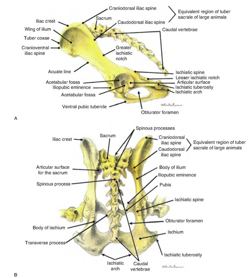
Fig. 6.27. Canine pelvic girdle. (A) Lateral view. (B) Dorsal view. (Reprinted from Constantinescu, 2002. With permission from the publisher.)
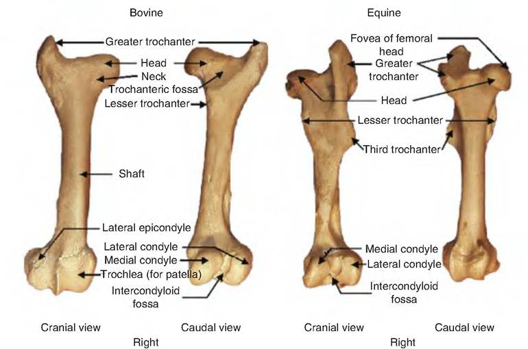
Fig. 6.28. Femur of bovine and equine.
with the other bones. The two hip bones are fused on the ventral midline at the pelvic symphysis. This fusion includes the two pubic and two ischial bones.
The ilium is the largest and most cranial of the os coxae, consisting of a wing and body. The ilium forms the cranial part of the acetabulum and articulates with the sacrum at the sacroiliac joint. In carnivores, the wing of the ilium is relatively unspecialized and its broad plane is oriented parallel with the longitudinal axis of the body. In large animals, the iliac wing is expanded and twisted so that the broad plane is oriented almost transverse to the longitudinal axis of the body. The modifications relate to increasing surface area for attachment of the powerful muscles of the hind limb in these larger and much heavier animals. The tuber coxae are the more lateral prominences of the ileac wings. Because the tuber coxae are particularly prominent in cattle, it is commonly referred to as the "hook." The tuber sacrale is the medial process of the wing that articulates with the sacrum. The ischium is the Caudalmost portion of the os coxae and forms the lateral portions of the obturator foramen, the large opening in the floor of each ox coxa. The ischiatic tuberosity is the caudalmost part of the ischium and is quite prominent. The ischiatic tuber is referred to as the "sit bones" in man and carnivores, and as the "pin bone" in cattle. The pubis forms the Cranioventral part of the os coxae. The pubis consists of a central body and two branches. The more medial branch contributes to the obturator for humans, while the more lateral branch contributes to the acetabulum.
The femur, or thigh bone, articulates proximally with the os coxae at the acetabulum, forming the hip joint, distally with the tibia forming the stifle (true knee) joint (Fig. 6.28). Asmall depression on the medial surface of the head of the femur, the fovea capitis, provides attachment for the round ligament of the femur. This ligament inserts in the acetabular fossa, anchoring the head of the femur into the acetabulum. The head of the femur continues with the body of the femor. The greater trochanter is the large prominence found lateral to the head of the femur; the lesser trochanter is the smaller prominence found distal to the head on the medial side. In horses, a prominent third trochanter also lies on the lateral side, distal to the greater trochanter. Note that trochanters are unique to the femur. The medial and lateral condyles are the two large rounded prominences at the distal end of the femur, and articulate with the tibia. Also on the distal end of the femur is the patellar surface, a groove bordered by two ridges that articulates with the patella. The patella, or kneecap, is the largest sesamoid bone of the body.
The tibia and fibula, or bones of the "lower leg" (crus), are located between the femur and metatarsal bones (Fig. 6.29). The tibia, or shin bone, is located medially, and is the weight-bearing bone of the crus. Located at the proximal end of the tibia are the medial and lateral condyles, separated by the intercondylar eminence. The tibia condyles articulate with the corresponding condyles of the femur. The fibula is located more laterally and is not weight bearing. Distally, the
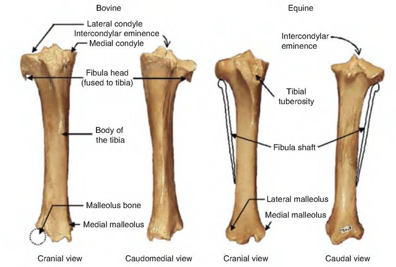
Fig. 6.29. Tibia and fibula of various species. The fibula for the equine was drawn to show where these bones would be if they were present on these specimens.
fibula articulates with the tibia and the fibular tarsal bone. The distal fibula in cattle is represented by the separate malleolar bone.
The tarsus, or "hock," consists of the three rows of bones between the crus and metatarsal region (Fig. 6.30). Similar to the carpus, this region is characterized by multiple bones arranged in multiple rows. However, the hock has a more complex arrangement than the carpus, with a proximal row, a sort of intermediate bone, and then a distal row. Again, marked species variation is present in the number of individual bones present. In all species, the proximal row consists of two bones. Beyond that, generally, marked variations occur among species in fusions among different bones and the resulting number of separate bones. To simplify, carnivores and pigs have seven tarsal bones, horses have six, and ruminants have five separate tarsal bones.
In all species, one of the two largest bones of the tarsus is the talus or tibial tarsal bone. The talus is located dorsomedially and articulates with the tibia and fibula or its equivalent via its trochlea. The calcaneus or fibular tarsal bone is the other bone in the proximal row, just lateral to the talus. The calcanean tuberosity is a large process of the calcaneus acting as a lever for the common calcanean (Achilles) tendon, and is commonly called the point of the hock.
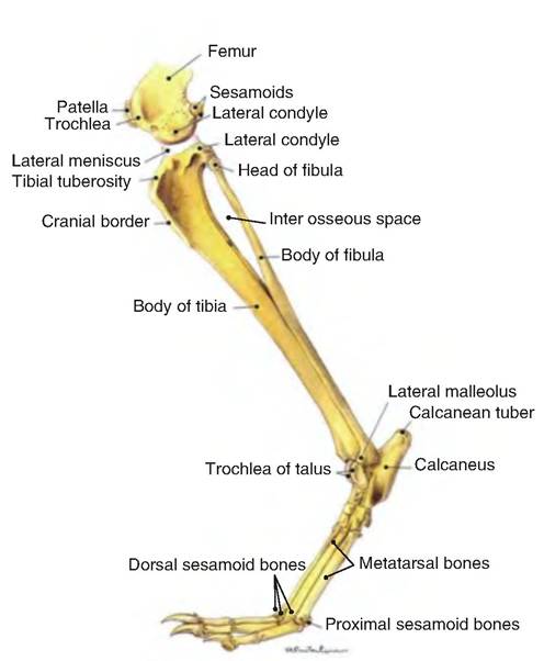
Fig. 6.30. Distal pelvic limb of the dog. The lateral aspect of the pelvic limb of the dog from the stifle to the toes. (Reprinted from Constantinescu, 2002. With permission from the publisher.)
Metatarsal bones and digits are located distal to the tarsus. In the horse and pig, they follow the same pattern as the thoracic limb. In carnivores, the first metatarsal bone is greatly reduced, and the digit is usually absent. Exceptions exist among certain breeds of dog, in which the dewclaw may be present or even doubled. Certain breed standards require the presence of hind dewclaws. Usually, these rear dewclaws are similar to the dewclaws of cattle, in that they contain only cutaneous (skin-related) and no bony elements. In ruminants, the first and fifth metatarsal bones are absent, and the second is reduced to a tiny element.
The digits of the pelvic limb follow the same pattern as in the thoracic limb: each weight-bearing digit is composed of three phalanges, with paired sesamoid bones on the plantar aspect of the metacarpophalangeal joint and a single distal sesamoid on the plantar aspect of the distal Interphalangeal joint.