Bones of the forelimb (Fig. 3.1)
The bones of the forelimb are:
Clavicle- frequently absent in the dog. When present, it is just a remnant of bone that lies in the muscles cranial to the shoulder joint - it is described as being vestigial.
T he clavicle is normally present in thecal but does not articulate with other bones.Scnpiilii - also called the shoulder blade (Fig. 3.14). ft is a large flat lx>ne found on the lateral surface of the trunk at the junction of the neck and ribs. It has a prominent ridge or spine running down the middle of its lateral surface. This divides the lateral surface into two regions: the supraspinous fossa and infraspinous fossa. On the distal end of the spine there is a bony projection called the acromion. At the
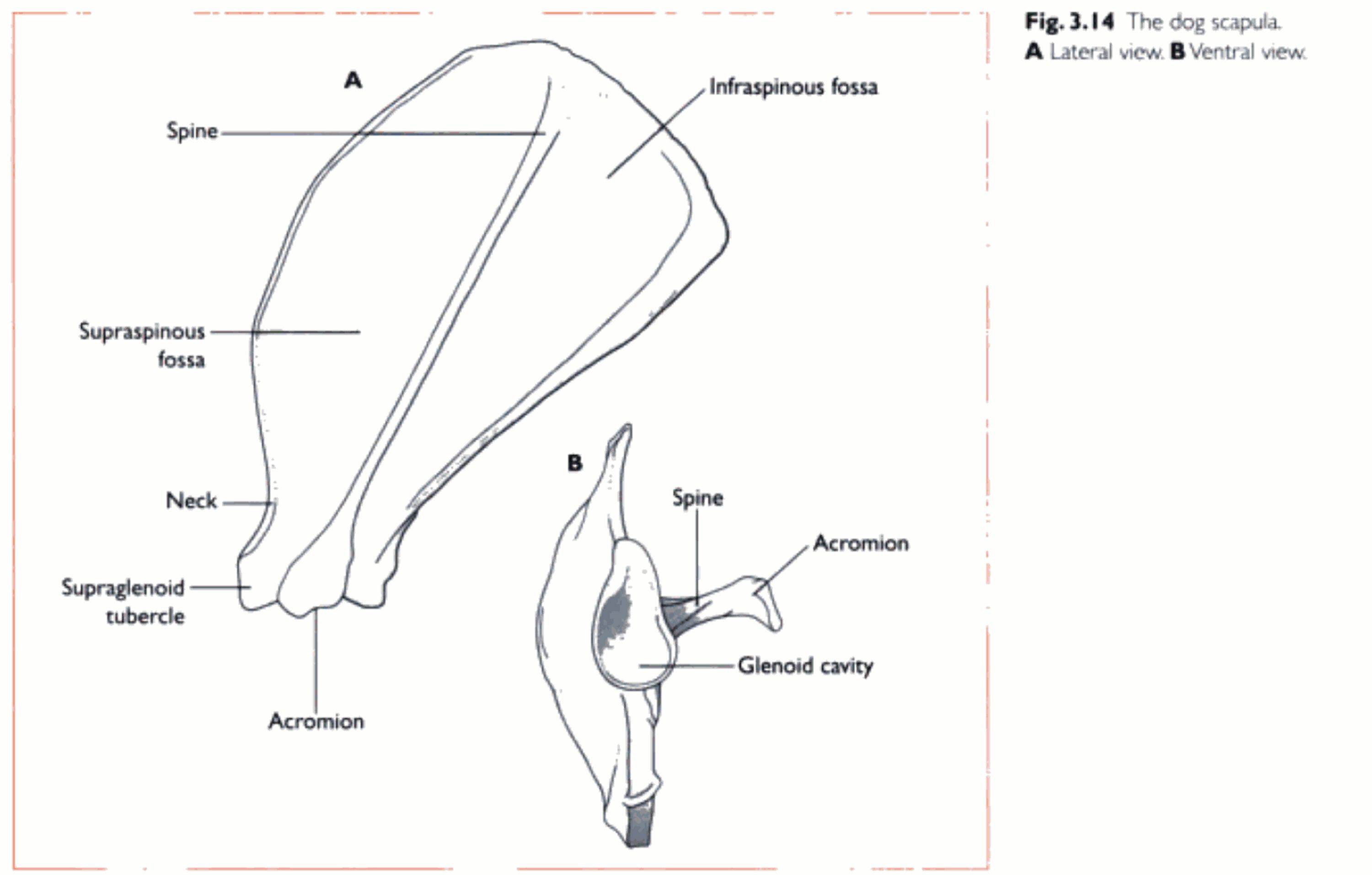
distal end of the scapula the bone narrows al the neck and there is a shallow articular socket, called the ylenoid cavity. which forms the shoulder joint with the head of the humerus. The medial surface of the scapula is Ilat and comparatively smooth. Humeriis - this a long bone forming the upper forelimb (Fig. 3.1 5). It articulates proximally with the scapula at the shoulder joint, and distally with the radius and ulna at (he elbow joint. The proximal end of the humerus consists of a large rounded projection, the head. Cranial and lateral to the head there is a large prominence, called the ιfreater tuhen le. Another prominence, the lesser tubercle. lies medial to the head. Both of these are sites for attachment of the muscles that support the shoulder joint. Distal to the head is the ne(see Fig. 3.2 3. p. 4 31. The talus, or tibial tarsal bone, is the most medial and has a proximal trochlea, which is shaped to lit the end of the tibia. The calcaneus, or Iibular Iarsal bone, is positioned laterally and has a Iargecaudal projection known as the tuber calcis. which forms the ‘point' of the hock.
Metatarsus and digits - these closely resemble the pattern of the metacarpus and digits in the forepaw. The metatarsus is composed of four metatarsal bones, although some breeds possess live, having a small metatarsal I or hind dew claw.
Fig. 3.20 Tne dog tve on the ventral surface of the os penis in the dog. In the cat the urethral groove is on the dorsal surface of the os penis, due to the different orientation of the penis (sec Ch. I 1).
The cow has a splanchnic bone in its heart, called the os coπlis. while the bird has splanchnic bones forming a rim around the eye to provide strength to the large eyeball Isee Ch. I J I.