CASES
CASE 1
Signalment/history
A 15-year-old, spayed female domestic shorthair cat presented for a mass in the left mandibular area. FNAs of the mass were submitted for cytologic evaluation.
Cytologic description
The sample was highly cellular with low numbers of erythrocytes and neutrophils in the background. The majority of cells were large rounded cells with abundant basophilic cytoplasm, a large round nucleus, stippled chromatin, and one to two large prominent nucleoli (Figures 4.68a, b). Very large ‘owl’s eye’ nucleoli were present in several cells. Marked anisocytosis and anisokaryosis were observed. There were several binucleate cells and aberrant mitotic figures. Low numbers of cells contained small black pigment consistent with melanin (Figure 4.68c).
Interpretation: Amelanotic melanoma.
Discussion
Amelanotic melanomas can be difficult to diagnose unless a few melanin granules can be found. This diagnosis should be suspected in cases where it is difficult to determine if the neoplastic cells are round, epithelial, or mesenchymal cells.
Case 2
Signalment/history
A 1-year-old, spayed female German Shorthair Pointer presented with a crusty skin lesion. A skin scraping was performed and submitted for cytologic evaluation.
Cytologic description
Slides were highly cellular with minimal peripheral blood contamination. They contained a mixed inflammatory cell population predominated by degenerate neutrophils with fewer macrophages and eosinophils. Small 3–6 μm diameter round deeply basophilic yeast-like structures were present individually and in clusters. Some septate branching hyphal structures were also observed (Figure 4.69a). Fungal organisms were seen extracellularly and within macrophages (Figures 4.69b, c). Rarely, short rod-shaped bacterial organisms were seen within neutrophils (Figure 4.69d).
Many mature squamous epithelial cells were present (Figure 4.69e). The background of the sample consisted of moderate cellular debris and basophilic proteinaceous material.Interpretation: Marked septic mixed inflammation with evidence of fungal (most suggestive of a dermatophyte) and bacterial organisms.
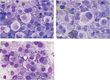
Figure 4.68a–c FNA of a mass in the left mandibular area of a cat. There are sheets of large rounded cells with abundant basophilic cytoplasm, a large round nucleus, stippled chromatin, and one to two large prominent nucleoli. ‘Owl’s eye’ nucleoli are present in several cells. Marked anisocytosis and anisokaryosis are observed. There are a few binucleate cells. (b,c) Low numbers of cells contain black pigment consistent with melanin. (Wright–Giemsa, 1,000? magnification.)
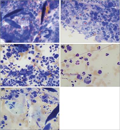
Figures 4.69a–e Skin scraping of a crusted lesion from a dog. The sample contains a mixed inflammatory cell population predominated by degenerate neutrophils with fewer macrophages and eosinophils. Small 3–6 μm diameter round deeply basophilic yeast-like structures are present individually and in clusters (red arrows). (a) A septate branching hyphal structure is shown (black arrow). (b, c) Fungal organisms are seen extracellularly and within macrophages. (d) Rarely, short rod-shaped bacterial organisms are observed within neutrophils (yellow arrow). (e) Many mature squamous epithelial cells are present (black stars; Wright–Giemsa, 1,000? magnification).
Discussion
Unstained slides were submitted for DNA isolation and polymerase chain reaction to determine the identity of the fungal organisms. The DNA sequence had 99% homology with Trichophyton mentagrophytes.
CASE 3
Signalment/history
A 3-year-old, female, domestic shorthair cat (indoor/outdoor) presented for a swollen nose and multiple, variably sized, small, hairless masses on both ears.
An FNA of one of the ear masses was submitted for cytological evaluation.Cytologic description
The slides were highly cellular and consisted of nucleated cells present in a background containing high numbers of red blood cells, low amounts of nuclear debris, and high numbers of small extracellular organisms (Figures 4.70a–c). There were high numbers of macrophages and lower numbers of neutrophils, and many contained low to high numbers of the same organisms. These were round to oval, approximately 2–3 μm in diameter, and contained a round purple-staining nucleus and a perpendicularly oriented small rod-shaped kinetoplast (Figure 4.70b). The organisms were also sometimes present in large variably sized phagocytic vacuoles within the macrophages, and were consistent with Leishmania amastigotes. Low numbers of bi-nucleated macrophages and multinucleated giant cells were also observed.
Interpretation: Marked mixed inflammation (predominantly macrophagic) with Leishmania amastigotes present.
Discussion
Unstained slides were submitted for PCR and DNA sequencing analysis to determine the species of Leishmania present. Results revealed the presence of Leishmania amazonensis.
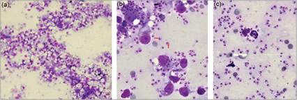
Figures 4.70a–c FNA of ear masses from a cat. The sample contains high numbers of extracellular and intracellular, small, round to oval organisms and high numbers of a mixture of inflammatory cells, with macrophages predominating (a). The organisms are round to oval, 2–3 μm in diameter, have an eccentrically located, round, purple staining nucleus and a small rod-shaped kinetoplast that has a perpendicular orientation (b, red arrows). They are present intracellularly within macrophages (b) and neutrophils (c), and extracellularly in very high numbers (Wright–Giemsa 200? magnification, A; 1,000? magnification, b and c). (a) High numbers of inflammatory cells (predominantly macrophages) and numerous extracellular and intracellular small, round to oval organisms. (b) The organisms are present extracellularly and within macrophages. The organisms contain an eccentrically located nucleus and a small rod-shaped kinetoplast that has a perpendicular orientation (red arrows). (c) Organisms are present extracellularly and within neutrophils.