Melanomas
Melanomas originate from melanocytes in the skin and can occur in the skin, mucosa, and even the anal sac. The morphology of melanocytes can be spindle-shaped or round with a round to variably shaped nucleus.
Cells may be individualized or aggregated on the slide. The distinguishing characteristic of melanocytes is the presence of round or rice-shaped, brown–black to blue–gray, melanin granules (Figures 4.66a, b). Small, well-circumscribed, well-differentiated, melanotic melanomas (sometimes referred to as melanocytomas) typically are benign in cats and dogs unless they are near a mucous membrane or a nail bed. Amelanotic melanomas are much more difficult to diagnose; because of the pleomorphism of melanocytes, there is virtually no morphologic indicator of cell type unless melanin granules are seen. Immunocytochemical stains, such as S-100 or Melan-A, can aid in making a diagnosis of amelanotic melanoma (Smith et al., 2002). Malignant melanomas, including most amelanotic melanomas, are very aggressive tumors that rapidly spread to draining lymph nodes. Cells tend to be large and pleomorphic with several criteria of malignancy. They may contain a somewhat distinctive, large, round, prominent nucleolus that takes up the majority of the nucleus, termed ‘owl’s eye’ nucleoli (Figure 4.67). Aspiration of draining lymph nodes and thoracic imaging is recommended whenever a malignant melanoma is diagnosed. 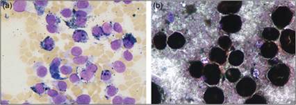
Figures 4.66a,b Melanomas. (a) FNA of a 1-cm black mass on the right pinna of a 4-year-old Labrador Retriever. The sample contained several individualized rounded to spindle-shaped cells with scant lightly basophilic cytoplasm, variable numbers of pinpoint black cytoplasmic granules, and a large round to ovoid nucleus with finely stippled chromatin. (b) FNA of a mass from an adult domestic shorthair cat. Cells in this sample are rounded and filled with black granules that obscure the morphology of the nucleus. Large numbers of black granules are present in the background of the sample (Wright–Giemsa, 1,000? magnification).
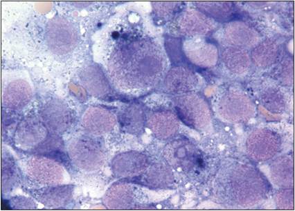
Figure 4.67 FNA of a dermal mass from a dog. Malignant melanocytes exhibit marked anisocytosis and anisokaryosis. They have a variable amount of lightly basophilic cytoplasm and an ovoid nucleus with stippled chromatin and a prominent nucleolus. A few variably sized black granules are seen in some of the cells. The cell at the top center of the image has a very large ‘owl’s eye’ nucleolus (Wright–Giemsa stain, 1,000? magnification).