CASES
CASE 1
Signalment/history
An 11-year-old spayed female Labrador Retriever. The animal presented to the orthopedic surgery service for a 12-week recheck after a tibial plateau leveling osteotomy (TPLO) to correct a ruptured cruciate ligament.
She was reportedly doing well at home, with minimal evidence of orthopedic pain.Clinical examination
The examining clinician noticed a 3-cm, freely movable, subcutaneous mass on the left side of the neck, in the area of the left submandibular lymph node. Three aspirates were obtained and submitted for cytologic examination.
Cytologic description
One of the aspirates contained a heterogeneous lymphoid population, composed primarily of small lymphocytes, with fewer medium and large lymphocytes, and rare plasma cells. This aspirate was most compatible with aspiration of a cytologically unremarkable lymph node (Figure 6.38). The other two aspirates contained a large amount of blood, which aligned in a ‘windrowing’ pattern. Occasional individualized foamy round cells, interpreted as either macrophages or individualized secretory epithelial cells, were observed. Low numbers of angular yellow hematoidin crystals, indicative of prior hemorrhage, were also noted within the cytoplasm of low numbers of these cells. Free in the background were abundant amorphous collections of eosinophilic to amphophilic material, cytologically most compatible with saliva.
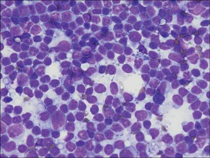
Figure 6.38 Lymph node. A heterogeneous lymphoid population, composed primarily of small lymphocytes with fewer medium and large lymphocytes. A small amount of hemosiderin and abundant lymphoglandular bodies are present in the background (modified Wright’s, 500? magnification).
Cytologic interpretation
The overall cytomorphology of this lesion was most compatible with a sialocele with previous hemorrhage (Figure 6.39).
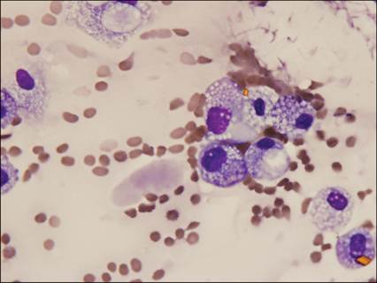
Figure 6.39 Facial swelling. Free blood in the background forms occasional linear structures (‘windrows’). Individualized to very loosely cohesive round shaped cells (either macrophages or secretory epithelial cells) are present. Two ‘mustard yellow’ rhomboid hematoidin crystals are shown. Morphologically compatible with a sialocele (modified Wright’s, 1,000? magnification).
Management
The owner elected to pursue surgical removal of the lesion, as repeated drainage simply resulted in lesion recurrence. Histologic examination of the architecture revealed abundant amounts of granulation tissue extending from the superficial dermis to the subcutis, which displaced adjacent adnexa. A focally extensive area of pyogranulomatous inflammation was admixed with abundant amounts of amphophilic, flocculent material (saliva) and cellular debris. Scattered lymphocytes and plasma cells and multifocal areas of hemorrhage were noted. A morphologic diagnosis of sialocele was made (Figure 6.40).
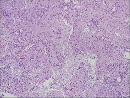
Figure 6.40 Histologic section of tissue from facial swelling. Plump, organizing fibroblasts (granulation tissue) surrounding a central mass of amphophilic flocculent material (saliva). Morphologic diagnosis: sialocele (H&E, 100? magnification).
Discussion
Sialoceles, also called salivary mucoceles, are accumulations of saliva in subcutaneous tissues with secondary purulent to pyogranulomatous inflammation. These lesions differ from ranulas (distensions of an epithelial lined salivary gland or duct) in that they are not lined by an epithelial lining. The exact cause of these lesions is unknown. However, it is likely that sialoceles are secondary to trauma, with disruption of a salivary duct resulting in leakage and encapsulation of saliva by connective tissue (Hostetter, 2023).
At the time of this patient’s TPLO surgery, 12 weeks prior to presentation, there was reportedly a malfunctioning pulse oximeter used for surgical monitoring. This resulted in much manipulation of the tongue throughout the anesthetic procedure, which may have caused rupture of the duct of the sublingual salivary gland.
Case 2
Signalment/history
An 8-year-old, neutered male Newfoundland presented for hematemesis and weakness. His owners brought in a tennis ball-sized structure that they found in his vomitus and believed to be a rubber ball.
Physical examination
Closer examination of the structure revealed that it was a solid piece of tissue. The tissue was bisected, and impression smears were made and submitted for cytologic evaluation. The patient was resistant to oral examination, and the owners elected not to sedate the animal to allow for proper restraint.
Cytologic/histologic evaluation
The impression smears were highly cellular and revealed a population of pleomorphic round-shaped to spindloid cells with abundant black, intracytoplasmic pigment granules. Nuclei were round to ovoid with coarsely granular chromatin and an occasional indistinct nucleolus. Low numbers of binucleated cells were seen. These findings were compatible with a cytologic diagnosis of melanoma (Figure 6.41). Scattered keratinized, anucleate squamous epithelial cells with adherent Conchiformibius spp. organisms were noted. The exact origin of the tumor was uncertain.
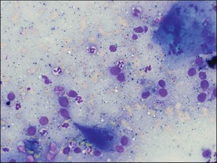
Figure 6.41 Scattered, individualized, round-shaped to spindloid cells are present with high N:C ratios and a scant amount of lightly basophilic cytoplasm. Nuclei are round to ovoid. Abundant black melanin granules are free in the background. Occasional deeply basophilic, anucleate squamous epithelial cells are present with adherent Conchiformibius spp. organisms, indicative of origin or contamination from the oral cavity.
Morphologically most compatible with melanoma (modified Wright’s, 500? magnification).
The tissue from which the impression smears were made was submitted for histologic examination (Figure 6.42). Histologic examination revealed a poorly demarcated, unencapsulated, infiltrative, densely cellular neoplasm composed of neoplastic melanocytes. Cells were arranged in sheets and interlacing streams supported by abundant fibrovascular stroma. Individual cells were pleomorphic, ranging from round to polygonal to spindloid, with variably distinct cell borders and scant to moderate amounts of eosinophilic cytoplasm containing variable numbers of melanin granules. Nuclei were centrally placed, ovoid to elongate, with vesiculated chromatin and one distinct, eosinophilic nucleolus. There were occasional karyomegaly and frequent binucleation. Anisocytosis and anisokaryosis were marked. There were 3 mitoses per 10 microscopic fields at 400? magnification. The melanoma was covered by moderately hyperplastic squamous epithelium with moderate parakeratotic hyperkeratosis, suggestive of gingival, buccal, or palatine origin.
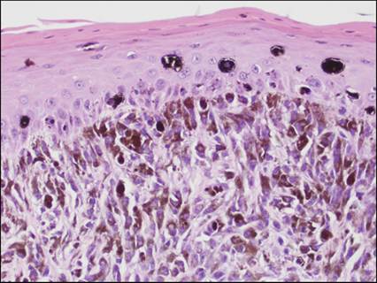
Figure 6.42 Streams of spindloid to polygonal cells with abundant brown intracytoplasmic pigment granules dorsally displace the epithelium, which is covered with parakeratotic squamous epithelium. Morphologic diagnosis: melanoma (H&E, 400 magnification).
Management/outcome
On receiving a diagnosis of melanoma, the owners allowed sedation, and oral examination revealed a large, ulcerated mass emanating from the soft palate. It is believed that the ‘vomited’ portion of the mass came from this location and that the dog was likely vomiting due to nausea induced by swallowing large amounts of blood arising from the tumor. Surgical debulking was attempted, and complete margins were unattainable. The patient died within 2 weeks of diagnosis.
CASE 3
Signalment/history
A 10-year-old neutered male domestic short-haired cat presented with a history of excessive salivation.
Clinical examination
The examining clinician noted a large, ulcerated mass beneath the tongue. Aspirates were obtained with sedation.
Cytologic description
The aspirates contained a pleomorphic population of individualized to minimally cohesive squamous epithelial cells, in varying stages of keratinization. Individual cells were large and round-shaped to angular, with abundant cytoplasm, often containing a large perinuclear clearing, typical of squamous epithelium. Occasional mitotic figures were noted. Anisocytosis was marked, with mild to moderate anisokaryosis. The number of neutrophils was increased over the amount of blood (Figure 6.43).

Figure 6.43 Sublingual mass. A pleomorphic population of individualized to minimally cohesive squamous epithelial cells in varying stages of keratinization, which exhibit marked anisocytosis and mild to moderate anisokaryosis. The number of neutrophils is increased over the amount of blood. (Courtesy Dr. Anne Barger.)
Cytologic interpretation
This lesion is consistent with a squamous cell carcinoma with neutrophilic inflammation. After biopsy, the diagnosis was confirmed with histologic examination of tissue architecture.
Management/outcome
The owners elected palliative care and euthanasia was elected one month after diagnosis, due to progression of symptoms and diminished quality of life.