Cases
CASE 1
Signalment/history
A 7-year-old, neutered male Standard Poodle presented for a 5-day history of hematuria and dysuria.
Physical examination
Physical examination was normal except for an ulcerated hemorrhagic mass on the penis.
FNAs of the mass were submitted for cytologic examination (Figures 11.60, 11.61; Bolfer et al., 2015). 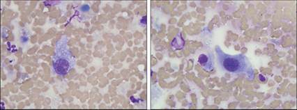
Figures 11.60,11.61 Hemangiosarcoma. FNA from a mass on the penis of a 7-year-old, neutered male Standard Poodle. Individualized large spindle-shaped cells with abundant basophilic cytoplasm, often several cytoplasmic vacuoles, and a large ovoid nucleus are seen (Wright–Giemsa, 1,000? magnification).
Cytologic description
The sample contained moderate numbers of large spindloid cells with deeply basophilic cytoplasm and an ovoid nucleus. Many cells contained punctate clear cytoplasmic vacuoles. Eosinophilic product was seen in low numbers of cells. Multinucleation, anisocytosis, anisokaryosis, and multiple prominent variably sized nucleoli were observed.
Cytologic interpretation
Sarcoma. Primary differential diagnoses are hemangiosarcoma and fibrosarcoma.
Management/outcome
Penile amputation was performed and the mass was submitted for histopathology. The histologic diagnosis was completely excised hemangiosarcoma. Two hundred and five days after the surgery, the patient presented to an emergency clinic with ascites and was euthanized due to presumptive disseminated hemangiosarcoma.
CASE 2
Signalment/history
A 10-year-old, spayed female Golden Retriever presented with a 2-month history of mucinous vaginal discharge.
Physical examination
No physical abnormalities were detected. A vaginal swab was submitted for cytologic examination (Figure 11.62).
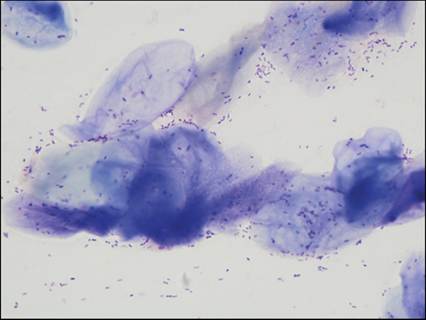
Figure 11.62 Possible estrus. Vaginal swab from a 10-year-old, spayed female Golden Retriever. The majority of cells are anucleate squamous epithelial cells. Large numbers of bacterial cocci and rods are observed in the background, but no inflammatory cells are seen (Wright–Giemsa, 1,000? magnification).
Cytologic description
The majority of cells were anucleate squamous epithelial cells with fewer nucleated squamous epithelial cells. Large numbers of bacterial cocci and rods were observed in the background. No inflammatory cells were seen.
Cytologic interpretation
Mature squamous epithelium. The bacteria are likely commensal organisms. If the sample is representative of the vaginal wall, not inadvertently sampled from the vulva or clitoris, it is consistent with estrus. In a spayed female, this is commonly associated with a retained ovarian remnant. Given the patient’s age and duration of the vaginal discharge, exogenous sources of estrogen (including neoplasms) should be considered.
Management/follow-up
The patient was fully evaluated for mass lesions. None were found. The sample was suspected of being more representative of the vulva than the vaginal wall.
CASE 3
Signalment/history
A 10-year-old, male castrated American Pit Bull Terrier presented for swelling of the caudoventral abdomen and scrotum.
Physical examination
The patient was in moderate pain and was administered analgesics. He had pitting edema of the abdomen and scrotum. His popliteal lymph nodes were enlarged. Ultrasonography revealed medial iliac and inguinal lymphadenopathy, and a prostatic cyst. FNA of the scrotum was performed (Figures 11.63, 11.64).
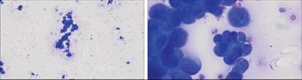
Figures 11.63,11.64 FNA from a scrotal swelling on a 10-year-old, male castrated Pit Bull.
Figure 11.63: Clusters of mesothelial cells are observed (Wright–Giemsa, 200? magnification). Figure 11.64: Mesothelial cells are present in clusters. They have a variable amount of round, deeply basophilic cytoplasm with a thin pink fringe at the cytoplasmic border. The nucleus is round or slightly cleaved and had coarse chromatin. One to two nucleoli are often seen. Anisocytosis and anisokaryosis are marked (Wright–Giemsa, 2,000? magnification).
Cytologic description
The sample was highly cellular. The majority of cells were clustered together. Most cells had a small amount of deeply basophilic cytoplasm, while others had more expanded, moderately basophilic cytoplasm. Cells frequently had a bright pink cytoplasmic fringe. The nucleus was round to ovoid with coarsely stippled chromatin and one to multiple distinct nucleoli. There were low numbers of binucleate cells. Marked anisocytosis and anisokaryosis were observed. Low numbers of vacuolated macrophages also were seen. No infectious organisms were present.
Cytologic interpretation
Probable malignant neoplasm. Differential diagnoses include reactive mesothelial tissue, mesothelioma, or an atypical carcinoma (possibly a prostatic or urogenital carcinoma).
Management/follow-up
Enlarged lymph nodes were aspirated for cytologic evaluation. The samples contained a reactive lymphoid population and cells similar to those observed in the swollen scrotum. The top differential diagnosis was a metastatic mesothelioma. Due to the pain the patient was in and the poor disease prognosis, the dog was euthanized.